Purdue-designed tool helps guide brain cancer surgery
July 2, 2014
 |
|
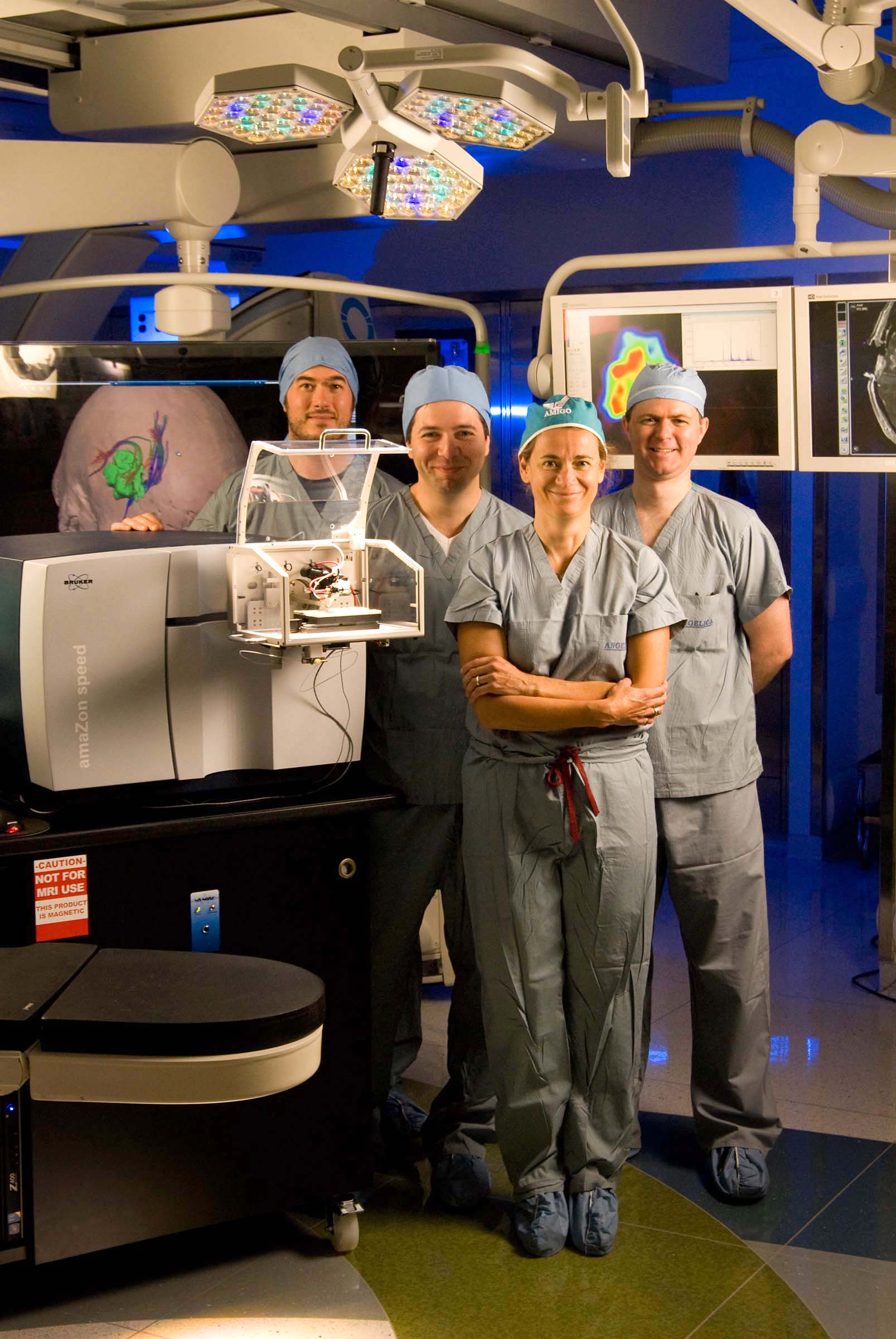
From left David Calligaris, Sandro Santagata Alexandra J. Golby and Isaiah Norton stand next to the Purdue-designed mass spectrometer in the Advanced Multi-Modality Image Guided Operating suite at Brigham and Women’s Hospital. (Photo courtesy of the Department of Neurosurgery, Brigham and Women’s Hospital, Boston) |
WEST LAFAYETTE, Ind. — A tool to help brain surgeons test and more precisely remove cancerous tissue was successfully used during surgery, according to a Purdue University and Brigham and Women's Hospital study.
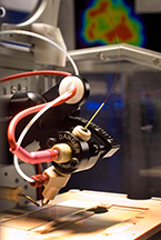
The Purdue-designed tool sprays a microscopic stream of charged solvent onto the tissue surface to gather information about its molecular makeup and produces a color-coded image that reveals the location, nature and concentration of tumor cells.
"In a matter of seconds this technique offers molecular information that can detect residual tumor that otherwise may have been left behind in the patient," said R. Graham Cooks, the Purdue professor who co-led the research team. "The instrumentation is relatively small and inexpensive and could easily be installed in operating rooms to aid neurosurgeons. This study shows the tremendous potential it has to enhance patient care."
Current surgical methods rely on the surgeon's trained eye with the help of an operating microscope and imaging from scans performed before surgery, Cooks said.
"Brain tumor tissue looks very similar to healthy brain tissue, and it is very difficult to determine where the tumor ends and the normal tissue begins," he said. "In the brain, millimeters of tissue can mean the difference between normal and impaired function. Molecular information beyond what a surgeon can see can help them precisely and comprehensively remove the cancer."
 |
|
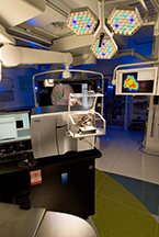
The Purdue-designed mass spectrometer to help guide brain surgery is shown in the Advanced Multi-Modality Image Guided Operating suite at Brigham and Women's Hospital. (Photo courtesy of Brigham and Women’s Hospital) |
The mass spectrometry-based tool had previously been shown to accurately identify the cancer type, grade and tumor margins of specimens removed during surgery based on an evaluation of the distribution and amounts of fatty substances called lipids within the tissue. This study took the analysis a step further by additionally evaluating a molecule associated with cell growth and differentiation that is considered a biomarker for certain types of brain cancer, he said.
"We were able to identify a single metabolite biomarker that provides information about tumor classification, genotype and the prognosis for the patient," said Cooks, the Henry Bohn Hass Distinguished Professor of Chemistry. "Through mass spectrometry all of this information can be obtained from a biopsy in a matter of minutes and without significantly interrupting the surgical procedure."
For this study, which included validation on samples and use during two patients’ surgical procedures, the tool was tuned to identify the lipid metabolite 2-hydroxyglutarate or 2-HG. This biomarker is associated with more than 70 percent of gliomas and can be used to classify the tumors, he said.
A paper detailing the results of the National Institutes of Health-funded study will be published in an upcoming issue of the Proceedings of the National Academy of Sciences and is published online.
In mass spectrometry molecules are electrically charged and turned into ions so that they can be identified by their mass. The new tool relies an ambient mass spectrometry analysis technique developed by Cooks and his colleagues called desorption electrospray ionization, or DESI, which eliminated the need for chemical manipulations of samples and containment in a vacuum chamber for ionization. DESI allows ionization to occur directly on surfaces outside of the mass spectrometers, making the process much simpler, faster and more applicable to surgical settings.
 |
|
The mass spectrometer portion of a Purdue-designed tool that assists brain surgeons in identification of tumor margins and cancerous tissue. (Photo courtesy of Brigham and Women's Hospital) |
The tool couples a DESI mass spectrometer with a software program designed by the research team that uses the results to characterize the brain tumors and detect boundaries between healthy and cancerous tissue. The program is based on earlier studies of lipid patterns that correspond to different types and grades of cancer and currently covers the two most common types of brain tumors, gliomas and meningiomas. These two types of tumors combined account for about 65 percent of all brain tumors and 80 percent of all malignant brain tumors, according to the American Brain Tumor Association.
Additional classification methodologies and metabolite biomarkers could be added to tailor the tool to different types of cancer, Cooks said.
The brain surgery was performed in the Advanced Multi-Modality Image Guided Operating suite, or AMIGO at Brigham and Women's Hospital.
Dr. Nathalie Agar, director of the Surgical Molecular Imaging Laboratory within the neurosurgery department at Brigham and Women's Hospital, led the study.
In addition to Cooks, co-authors of the paper from Purdue include former graduate student Livia S. Eberlin, who now is a postdoctoral researcher at Stanford University, and graduate student Joshua Wiley.
In addition to the National Institutes of Health, the James McDonnell Foundation, the Daniel E. Ponton Fund for the Neurosciences, the Sontag Foundation, the Ivy Foundation, the V Foundation, the Jared Branfman Sunflowers for Life Fund, the National Center for Image Guided Therapy, and a U.S. Army Medical Research grant funded this research.
Writer: Elizabeth K. Gardner, 765-494-2081, ekgardner@purdue.edu
Source: R. Graham Cooks, 765-494-5263, cooks@purdue.edu
Media contact for Brigham and Women’s Hospital: Jessica Maki, 617-525-6373, jmaki3@partners.org
Related news release:
New tool to help brain surgeons one step closer to operating room
ABSTRACT
Intraoperative mass spectrometry mapping of an onco-metabolite to guide brain tumor surgery
Sandro Santagata, Livia S. Eberlin, Isaiah Norton, Dvid Calligaris, Daniel R. Feldman, Jennifer L. Ide, Xiaohui Liu, Joshua S. Wiley, Matthew L. Vestal, Shakti Ramkissoon, Daniel A. Orringer, Kristen K. Gill, Ian F. Dunn, Dora Dias-Santagata, Keith L. Ligon, Ferenc A. Jolesz, Alexandra J. Golby, R. Graham Cooks, and Nathalie Y.R. Agar
For many intraoperative decisions, surgeons depend on frozen section pathology, a technique developed over 150 years ago. Technical innovations that permit rapid molecular characterization of tissue samples at the time of surgery are needed and in most cases, during the surgical procedure. Here, using desorption electrospray ionization mass spectrometry (DESI-MS), we rapidly detect the tumor metabolite 2-hydroxyglutarate (2-HG) from tissue sections of surgically-resected gliomas, under ambient conditions and without complex or time-consuming preparation. With DESI MS, we identify IDH1-mutant tumors with both high sensitivity and specificity within minutes, immediately providing critical diagnostic, prognostic and predictive information. Imaging tissue sections with DESI MS shows that the 2-HG levels correlate with tumor content, thereby indicating tumor margins. Mapping the 2-HG signal onto three-dimensional MRI reconstructions of tumors allows the integration of molecular and radiologic information for enhanced clinical decision-making. We also validate the methodology and its deployment in the operating room – we have installed a mass spectrometer in our Advanced Multimodality Image Guided Operating (AMIGO) suite and demonstrate the molecular analysis of surgical tissue during brain surgery. This work indicates that metabolite-imaging mass spectrometry could transform many aspects of surgical care.
