Listeria clever at finding its way into bloodstream, causing sickness

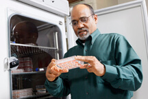
Arun Bhunia determined that listeria bacteria can pass between intestinal cells and triggers a mechanism that increases listeria's ability to enter the cells. (Purdue Agricultural Communication photo/Tom Campbell)
WEST LAFAYETTE, Ind. - Pathogenic listeria tricks intestinal cells into helping it pass through those cells to make people ill, and, if that doesn't work, the bacteria simply goes around the cells, according to a Purdue University study.
Arun Bhunia, a professor of food science, and Kristin Burkholder, a former Purdue graduate student who is now a postdoctoral researcher in microbiology and immunology at the University of Michigan Medical School, found that listeria, even in low doses, somehow triggers intestinal cells to express a new protein, heat shock protein 60, that acts as a receptor for listeria. This may allow the bacteria to enter the cells in the intestinal wall and exit into a person's bloodstream. Bhunia and Burkholder's findings were published in the early online version of the journal Infection and Immunity.
"It's possible that host cells generate more of these proteins in order to protect themselves during a stressful event such as infection," Burkholder said. "Our data suggest that listeria may benefit from this by actually using those proteins as receptors to enhance infection."
Listeria monocytogenes is a foodborne bacteria that can cause fever, muscle aches, nausea and diarrhea, as well as headaches, stiff neck, confusion, loss of balance and convulsions if it spreads to the nervous system. According to the U.S. Centers for Disease Control and Prevention, it sickens about 2,500 and kills 500 people each year in the United States and primarily affects pregnant women, newborns, older adults and those with weakened immune systems.
The findings suggest that listeria may pass between intestinal cells to sort of seep out of the intestines and into the bloodstream to cause infection.
"That can expedite the infection," Bhunia said.
Measurable increases of the heat shock 60 protein were detected when listeria was introduced to cultured intestinal cells.
Bhunia and Burkholder also introduced listeria to intestinal cells in the upper half of a dual-chamber container and counted the number of bacteria that passed through the cells and appeared in the lower chamber.
The bacteria moved to the lower chamber faster than it is known to do when moving through cells, and did so even when a mutant form of the bacteria that do not invade the intestinal cells was used. This suggests the bacteria are moving around the cells, Bhunia said.
"The infective dose is very low. Even 100 to 1,000 listeria cells can cause infection," Bhunia said. "We believe that these mechanisms are what allow listeria to cause infections at such low levels."
Bhunia said he next would try to understand how listeria and the heat shock 60 protein interact and work to develop methods to protect intestinal cells from the bacteria. The Center for Food Safety Engineering at Purdue funded part of the research.
Writer: Brian Wallheimer, 765-496-2050, bwallhei@purdue.edu
Sources: Arun Bhunia, 765-494-5443, bhunia@purdue.edu
Kristin Burkholder, 734-763-5639, kburkhol@umich.edu
Ag Communications: (765) 494-2722;
Keith Robinson, robins89@purdue.edu
Agriculture News Page
ABSTRACT
Listeria monocytogenes Uses LAP to Promote Bacterial Transepithelial Translocation and Induces Expression of LAP Receptor Hsp60
Kristin M. Burkholder and Arun K. Bhunia
Listeria monocytogenes interaction with the intestinal epithelium is a key step in infection process. We demonstrated that LAP promotes adhesion to intestinal epithelial cells and facilitates extraintestinal dissemination in vivo. The LAP receptor is a stress response protein Hsp60, but the precise role for the LAP-Hsp60 interaction during Listeria infection is unknown. Here we investigated the influence of physiological stressors and Listeria infection on host Hsp60 expression and LAP-mediated bacterial adhesion, invasion and transepithelial translocation in an enterocyte-like Caco-2 cell model. Stressors such as heat (41oC), TNF (100U), and L. monocytogenes infection (104-106 CFU/ml) significantly (P <0.05) increased plasma membrane and intracellular Hsp60 in Caco-2 cells, and consequently, enhanced LAP-mediated L. monocytogenes adhesion but not invasion to Caco-2 cells. In the transepithelial translocation experiment, WT exhibited 2.7-fold greater translocation through Caco-2 monolayers compared to lap-mutant suggesting LAP is involved in transepithelial translocation, potentially via a paracellular route. shRNA suppression of Hsp60 in Caco-2 reduced WT adhesion and translocation by 4.5- and 3-fold, respectively, while adhesion remained unchanged for the lap-mutant. Conversely, overexpression of Hsp60 in Caco-2 cells enhanced WT adhesion and transepithelial translocation, but not the lap-mutant. Further, initial infection with low dosage (106 CFU/ml) of L. monocytogenes increased plasma membrane and intracellular expression of Hsp60 significantly, which rendered Caco-2 cells more susceptible to subsequent LAP-mediated adhesion and translocation. Data provide insight into role of LAP as a virulence factor during intestinal epithelial infection, and pose new questions regarding the dynamics between the host stress response and pathogen infection.
