Purdue Sleep and Developmental Studies Lab uncovers potentially eye-opening link between ‘N2’ sleep, Alzheimer’s
A.J. Schwichtenberg
Written by: Tim Brouk, tbrouk@purdue.edu
New, potentially pioneering research in the Purdue University Sleep and Developmental Studies Laboratory is examining deep sleep’s role in “sloshing” metabolic waste from the brain.
The study led by A.J. Schwichtenberg, associate professor in the Purdue Department of Human Development and Family Science; her graduate student Moon West; Yunjie Tong, associate professor in the Weldon School of Biomedical Engineering; and his graduate student Andrew Frels has analyzed MRI and 36-channel electroencephalogram (EEG) scans of 33 adults ages 30-55 so far as they sleep. The researchers have found cerebrospinal fluid (CSF) flushes cellular brain waste earlier than originally thought — the waste gets cleared out via CSF sloshing during N2 or intermediate sleep in adults’ brains. Intermediate sleep occurs between light sleep (N1) and deep sleep (N3).
The findings could inform sleep’s role in better neurological health and keeping neurodegenerative diseases, such as Alzheimer’s, at bay.
“Previous studies have documented high levels of CSF movement in N3. We were the first lab to document that it also takes place in N2,” Schwichtenberg said. “We’re kind of on the front cusp of this knowledge wave.”
The collaboration between the College of Health and Human Sciences and the Weldon School of Biomedical Engineering began in fall 2024. Data are drawn from community participants between the ages of 30 and 55 who get hooked up with the EEG cap before a two-hour sleep session in the Purdue Life Science MRI Facility. If N3/deep sleep is achieved, the participants return for a daytime scan to undergo breathing exercises based on their deep sleep breathing patterns and other tasks that monitor brain activity. Their heart rates and respiration rates are also monitored.
The work is funded through a two-year, $500,000 R21 grant by the National Institute of Aging through the National Institutes of Health.
The study is seeking 50 adults in all. If you would like to participate, email sleep@purdue.edu.
Alzheimer’s link
Some of the brain chemicals that the waste consists of include amyloid-beta, a peptide and key component in amyloid plaques often found in the brains of people with Alzheimer’s disease, and tau tangles, abnormal accumulations of the tau protein that are also commonly found in Alzheimer’s patients’ brains. Moving chemical buildups in the brain via the flow of CSF could fight off potential for Alzheimer’s.
Schwichtenberg explained that chemicals like amyloid-beta and tau stem from the glymphatic system, a network of vessels in the brain that plays a pivotal role in clearing toxins from the central nervous system. This area of the brain is “very active when we sleep.”
“In particular, it’s really active during slow-wave sleep. It’s kind of our deepest sleep sometimes what we refer to as our restorative sleep,” she added. “And so, what we are looking at is how sleep relates to the movement of CSF. And the movement of CSF is a key component of the glymphatic system.”
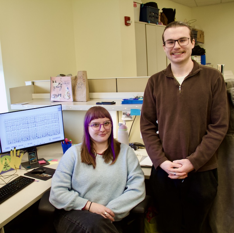
Graduate students Moon West, left, and Andrew Frels have teamed up to analyze thousands of data points produced by dozens of MRI and EEG scans.(Tim Brouk)
MRI and EEG working together
Schwichtenberg said the use of MRI is a novel approach to measure CSF movement in and out of the brain.
While it can be a challenge to get a participant to snooze for the entire two-hour window due to the EEG cap and the MRI noise, both methods are necessary to accurately measure CSF movement during the different sleep stages. So, to get participants to conk out, participants spent weeks prior acclimating themselves to the MRI noise by listening to similar sounds at home. They also adjust their sleep schedule, so they are deep in “sleep debt” by the time their sleep study is conducted.
Another arm of the work is to see if the sloshing occurs during an awake but rested — almost meditative — state. This means that adults could stave off Alzheimer’s without putting on their pajamas.
“We pull out their respiration patterns, and we have a code that we use in Python where a ball moves up and down (on a projection inside the MRI scanner). As the ball moves, they inhale and exhale,” West said. “What’s interesting is most people leave the (breathing exercise) scan and say, ‘I was about to fall asleep. I got so sleepy during it.’ It’s just the act of deep breathing and laying down and being relaxed.”
Tong’s lab specializes in MRI analysis. Frels said previous research experience has dealt with blood flow in the human body. Looking at CSF has similar attributes and methodology. The combined expertise between labs has had beneficial results in the study so far.
The MRI expertise couples well with the advanced MRI technology in the Purdue facility too. The scanner Schwichtenberg and the team are using is “multi-channel,” which means multiple coils are used to receive or transmit radiofrequency signals during an MRI scan. This approach enhances the signal-to-noise ratio, improves spatial resolution and allows for faster data acquisition.
“There’s only maybe six other labs in the U.S. that can even do that,” Schwichtenberg said. “We have a pretty unique setup here at Purdue that we can even capture all of those signals simultaneously.”
16,773 images per scan
West revealed the study was their first foray into dissecting large biosignal research data sets. The MRI and EEG scans offer a hefty amount of information as the brain sloshes during rest and sleep.
“It’s really cool to be able to look at people’s brains,” West said. “But my favorite part of it is cleaning and then scoring the EEG data. I never knew that you could tell how people were sleeping by their EEG data until this project. And it’s just really beautiful data.”
This data include 16,773 images per MRI scan. Most participants will have two scans. With 50 participants, the research team could be looking at more than 1.6 million images of human brain activity by study’s end.
“Right now, we’re working through pre-processing all the data because each scan is over a terabyte of data just sitting on our hard drive of raw files,” Frels explained. “Now, we have to push through all that to get it to a workable state where we can pull out information.”
This summer, the study will continue to scan participants as they sleep and conduct breathing exercises. While the final work won’t be completed until 2026, the team plans to publish early findings online later in 2025.
“The summer plan is to process all of this data,” Schwichtenberg said. “Regardless of what we find, we want the world to know about it and put that out there. We’ll be publishing on this data for five years.
“The richness of what people share with us will definitely help to inform the field.”
Discover more from News | College of Health and Human Sciences
Subscribe to get the latest posts sent to your email.