New imaging technology could reveal cellular secrets
April 25, 2013
 |
|
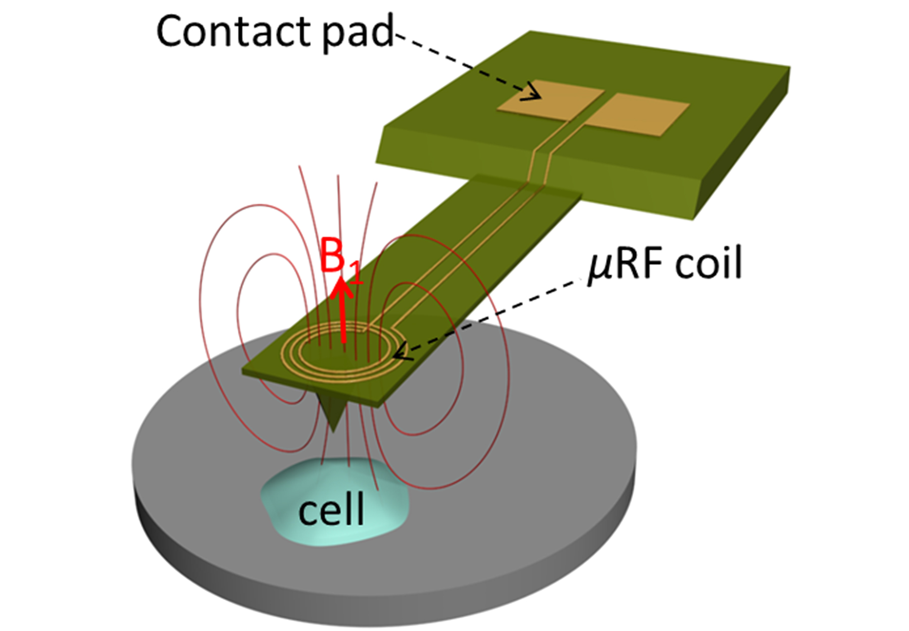
This image illustrates the concept for a new type of technology that combines two biological imaging methods - atomic force microscopy and nuclear magnetic resonance - to create a new way to study cancer-cell metastasis and other disease-related processes. (Purdue University image/ Xin Xu) |
WEST LAFAYETTE, Ind. - Researchers have married two biological imaging technologies, creating a new way to learn how good cells go bad.
"Let's say you have a large population of cells," said Corey Neu, an assistant professor in Purdue University's Weldon School of Biomedical Engineering. "Just one of them might metastasize or proliferate, forming a cancerous tumor. We need to understand what it is that gives rise to that one bad cell."
Such an advance makes it possible to simultaneously study the mechanical and biochemical behavior of cells, which could provide new insights into disease processes, said biomedical engineering postdoctoral fellow Charilaos "Harris" Mousoulis.
Being able to study a cell's internal workings in fine detail would likely yield insights into the physical and biochemical responses to its environment. The technology, which combines an atomic force microscope and nuclear magnetic resonance system, could help researchers study individual cancer cells, for example, to uncover mechanisms leading up to cancer metastasis for research and diagnostics.
The prototype's capabilities were demonstrated by taking nuclear magnetic resonance spectra of hydrogen atoms in water. Findings represent a proof of concept of the technology and are detailed in a research paper that appeared online April 11 in the journal Applied Physics Letters. The paper was co-authored by Mousoulis; research scientist Teimour Maleki; Babak Ziaie, a professor of electrical and computer engineering; and Neu.
"You could detect many different types of chemical elements, but in this case hydrogen is nice to detect because it's abundant," Neu said. "You could detect carbon, nitrogen and other elements to get more detailed information about specific biochemistry inside a cell."
An atomic force microscope (AFM) uses a tiny vibrating probe called a cantilever to yield information about materials and surfaces on the scale of nanometers, or billionths of a meter. Because the instrument enables scientists to "see" objects far smaller than possible using light microscopes, it could be ideal for studying molecules, cell membranes and other biological structures.
However, the AFM does not provide information about the biological and chemical properties of cells. So the researchers fabricated a metal microcoil on the AFM cantilever. An electrical current is passed though the coil, causing it to exchange electromagnetic radiation with protons in molecules within the cell and inducing another current in the coil, which is detected.
The Purdue researchers perform "mechanobiology" studies to learn how forces exerted on cells influence their behavior. In work focusing on osteoarthritis, their research includes the study of cartilage cells from the knee to learn how they interact with the complex matrix of structures and biochemistry between cells.
Future research might include studying cells in "microfluidic chambers" to test how they respond to specific drugs and environmental changes.
A U.S. patent application has been filed for the concept. The research has been funded by Purdue's Showalter Trust Fund and the National Institutes of Health.
Writer: Emil Venere, 765-494-4709, venere@purdue.edu
Sources: Corey Neu, 765-496-1426, cpneu@purdue.edu
Charilaos "Harris" Mousoulis, cmousoul@purdue.edu
Note to Journalists: Journalists can obtain a copy of the research paper by contacting Emil Venere, Purdue News Service, at 765-494-4709, venere@purdue.edu
ABSTRACT
Atomic Force Microscopy-Coupled Microcoils for Cellular-Scale Nuclear Magnetic Resonance Spectroscopy
Charilaos Mousoulis,1 Teimour Maleki,2 Babak Ziaie,1,2,3 and Corey P. Neu1,a
1 1Weldon School of Biomedical Engineering, Purdue University
2 2Department of Electrical and Computer Engineering, Purdue University
3 Birck Nanotechnology Center, Purdue University
We present the coupling of atomic force microscopy (AFM) and nuclear magnetic resonance (NMR) technologies to enable topographical, mechanical and chemical profiling of biological samples. Here, we fabricate and perform proof-of-concept testing of radiofrequency planar microcoils on commercial AFM cantilevers. The sensitive region of the coil was estimated to cover an approximate volume of 19.4 10 3 lm3 (19.4 pl). Functionality of the spectroscopic module of the prototype device is illustrated through the detection of 1H resonance in deionized water. The acquired spectra 14 depict combined NMR capability with AFM that may ultimately enable biophysical and biochemical studies at the single cell level. VC 2013 AIP Publishing LLC. [http://dx.doi.org/10.1063/1.4801318].
