Lab Culture: Uzay Emir Lab
By Tim Brouk, tbrouk@purdue.edu
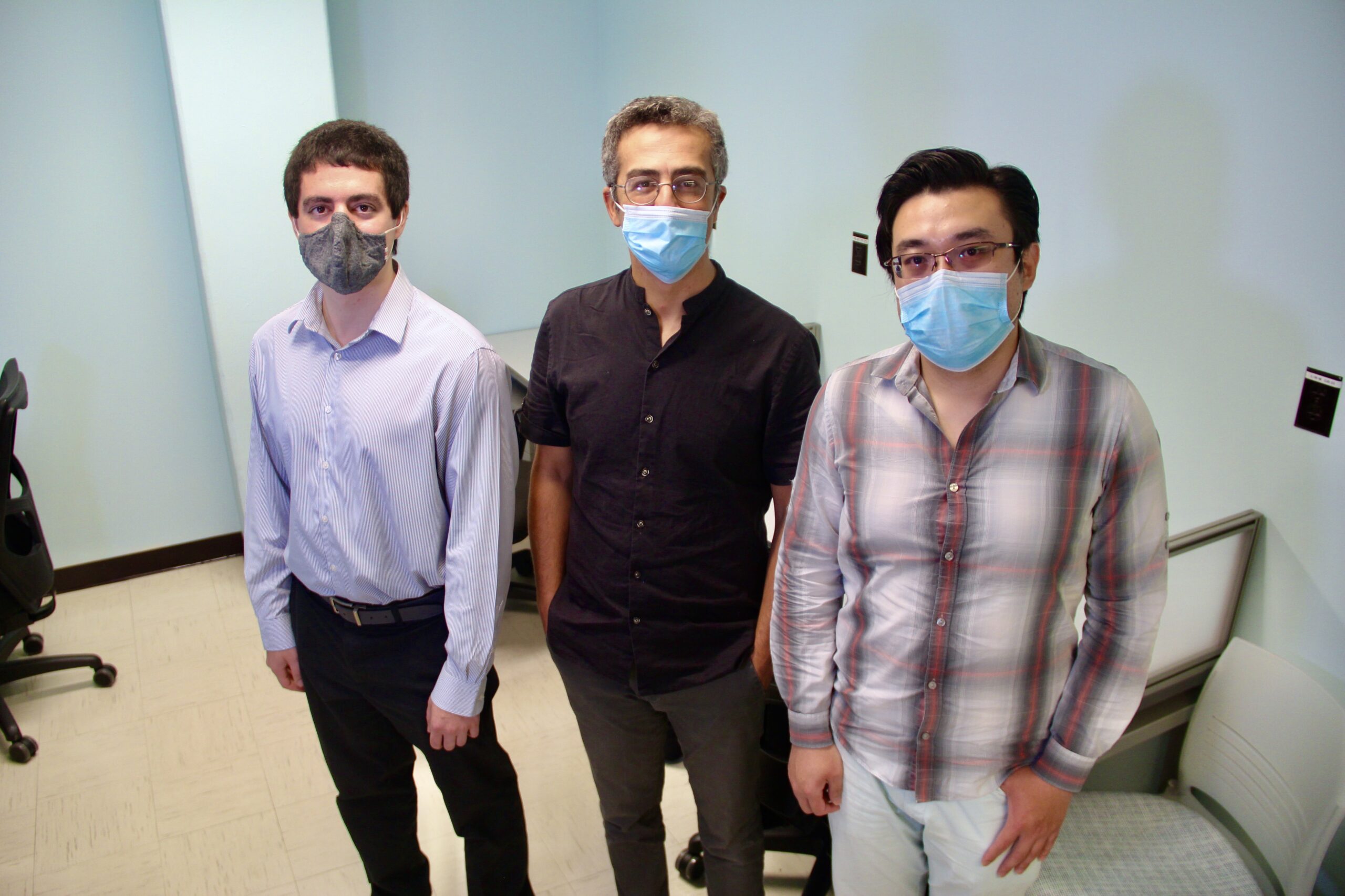
Health Sciences Assistant Professor Uzay Emir, middle, stands with his doctoral students Nicholas Farley, left, and Xin Shen inside their new lab space.Tim Brouk
One of School of Health Sciences Assistant Professor Uzay Emir’s imaging developments for detecting neurological disorders shares its name with a video conferencing software we’ve come to know all too well during the last 18 months.
In his lab and in the Purdue University Life Science MRI Facility, Emir’s work on his ZOOM magnetic resonance spectroscopic imaging (MRSI) technique published around the same time millions were learning to mute and unmute on Zoom from their laptops. Emir insisted his ZOOM name choice was pure coincidence.
“When COVID-19 started, we had submitted our first ZOOM MRSI paper. It was not intentional,” said Emir with a laugh. “Now everybody is Zooming so we assume ZOOM MRSI will be the next generation of spectroscopic imaging.”
Emir’s projects strive to expand and build upon MRSI, structural MRI and fMRI techniques. fMRI, or functional magnetic resonance imaging, measures blood flow and metabolic function while structural MRI, or magnetic resonance imaging, examines bones and organs in the body.
Complementary to these, magnetic resonance spectroscopy imaging (MRSI) techniques can be utilized to reveal abnormalities at the molecular level before any visible macroscopic changes in anatomy and physiology occur.
MRSI provides unique information on the chemical composition distribution of the human tissue non-invasively. The technique is based on the same physical principles as MRI. However, the MRSI measurement results in a spectrum where metabolite nuclei resonate in characteristic frequencies, depending on their chemical and physical vicinity in a molecule. The surroundings change the local magnetic field experienced by the nuclei, and thus their resonance frequency can be measured.
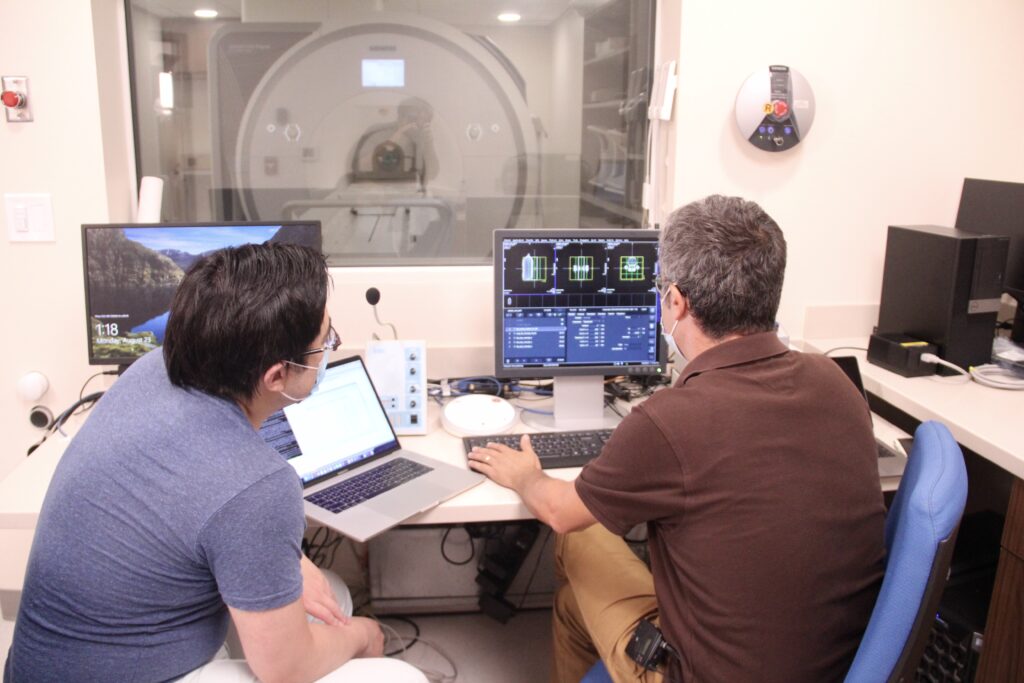
Shen and Emir prepare for an MRI scanning experiment. 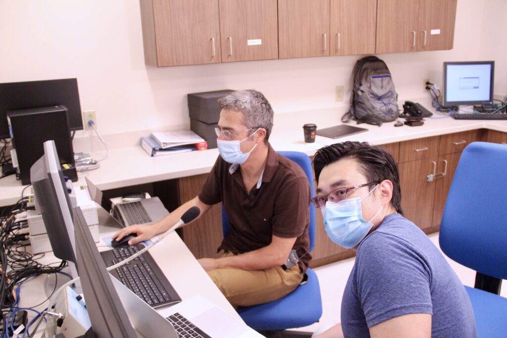
Lately, Emir’s research focuses on obtaining molecular images in the brain at the highest possible resolution as well as understanding iron and myelin content within the brain and how it relates to autism spectrum disorder (ASD).
“We assume that there are some molecules in the human brain that differ in kids with ASD compared to the typically developed ones,” Emir said. “And that might be reflected as iron and myelin content alterations along with neurotransmitters such as glutamate and GABA (gamma aminobutyric acid).”
The researcher recently applied for ZOOM MRSI patents.
“We want to get ZOOM on our scanners and scanners around the world to investigate with more detail,” said Emir, adding that this work has expanded to detection of diseases in other parts of the body, such as kidney cancer, liver disease and hypertension.
Calculate then scan
Doctoral student Xin Shen created 70,000 calculations for an imaging project that looks at how cartilage reacts to physical stress by imaging the sodium content in the body. The countless hours of computation were done in Emir’s new lab space in Hampton Hall before initial testing began in the MRI Facility. The lab in Hampton is primarily used for software coding, simulations and meetings.
Emir and his students did the paint work themselves, opting for a light blue. Emir obtained the room right before the COVID-19 pandemic forced remote work. This summer, he was happy to open it up again and get back to it.
When projects are taken to the MRI Facility for testing, the researchers set up multiple computers in a control room sealed away from powerful MRI scanners. Signs on a heavy door warn of a “strong magnetic field,” while Emir and his students monitor the experiments through a window. Communication is done through an intercom patched into a participant’s headphones.
A recent visit saw the researchers testing Shen’s technique on bottles of salt water. Eventually, human participants will replace the bottles.
“If we have the highest resolution, we can quantify important features in the cartilage to prevent future injuries,” Emir said. “The concentrations of sodium are quite high and allow us to detect.”
Netflix ‘n’ scan?
Since early detection of ASD could lead to better treatment plans and easier lives, children are often scanned for Emir’s work in the MRI Facility.
To keep them calm and still, Emir installed a projector near the MRI machine. A movie or TV show is projected in reverse before it is reflected from a mirror into the MRI machine, where the young patient can watch their favorite Disney movie. Emir patches in a laptop loaded with all the video streaming services as well as Spotify. Audio of the movies, music and instructions are piped from the control room into the participant’s headphones. Emir prefers when participants listen to music during the scan, as some fall asleep, making it easier for him to get more accurate imaging.
A large container of earplugs waits for participants, as the MRI machine is loud. Most scans take less than an hour, but laying still in such a setting is stressful. Emir encourages all his students to take a test scan to know what it’s like to be in an MRI machine.
“They need to understand what’s happening to the subjects during the scan,” Emir said. “The participants are doing a huge amount of the work, being in the scanner and helping our research. If we don’t have them and their willingness, then we can’t do our research. It would never go to the translational level.”
On the ’Tube
Emir has made it a practice to upload simulations, experiment results and PowerPoint presentations on his research to YouTube. His channel is home to 25 videos that explore the possibilities of imaging techniques. The videos explain the research as well as promote it.
“Whatever we develop before going to publication, we always upload results to the YouTube channel,” Emir said. “We want to let people know what we’re working on. We want the public engagement and awareness of what we are working on. We want to show our successes before they are published.”
That success attracted graduate students like Shen to Emir’s lab. Fellow doctoral student Nicholas Farley found a “natural” niche in Emir’s research, bringing a passion for MRI and other types of imaging.
“This is a very nice lab to work in, and I’ve enjoyed it since the first day. You know that you’re helping the community at large by potentially being a part of something bigger than yourself in creating these sequences, which might be able to help everyone out in the future,” he explained. “MRI and studying metabolites in the brain can be allowed to diagnose things before they actually happen. Being a part of this is a way to give back to the community, and it always feels great to pursue scientific advancements. It’s a great feeling to help out here.”
Emir appreciates the many hours his students put into his lab, and he looks forward to collaborating with them after graduation.
“Students that are working with me will be my colleagues at the end of this journey,” Emir said. “We will have hard times, easy times, happiness, sadness and difficulties, but we survive. I want my future colleagues to be successful.”
Discover more from News | College of Health and Human Sciences
Subscribe to get the latest posts sent to your email.