
| VIDEO |
 * Animation of the crystal (09 seconds) * Animation of the crystal (09 seconds) |
![]()
![]()
January 2, 2008
Scientists find missing evolutionary link using tiny fungus crystal
WEST LAFAYETTE, Ind. - |
By studying the three-dimensional version of the fungus protein bound to an RNA molecule, scientists from Purdue University and the University of Texas at Austin have been able to visualize how life progressed from an early self-replicating molecule that also performed chemical reactions to one in which proteins assumed some of the work.
"Now we can see how RNA progressed to share functions with proteins," said Alan Lambowitz, director of the University of Texas Institute for Cellular and Molecular Biology. "This was a critical missing step."
Results of the study were published in Thursday's (Jan. 3) issue of the journal Nature.
"It's thought that RNA, or a molecule like it, may have been among the first molecules of life, both carrying genetic code that can be transmitted from generation to generation and folding into structures so these molecules could work inside cells," said Purdue structural biologist Barbara Golden. "At some point, RNA evolved and became capable of making proteins. At that point, proteins started taking over roles that RNA played previously - acting as catalysts and building structures in cells."
 |
In order to show this and learn more about the evolution from RNA to more complex life forms, Lambowitz and Paul Paukstelis, lead author and a research scientist at the Texas institute, needed to be able to see how the fungus' protein worked. That's where Golden's team joined the effort and crystallized the molecule at Purdue's macromolecular crystallization facility.
"Obviously, we can't see the process of moving from RNA to RNA and proteins and then to DNA, without a time machine," Golden said. "But by using this fungus protein, we can see this process occurring in modern life."
Looking at the crystal, the scientists saw two things, Golden said. One was that this protein uses two completely different molecular surfaces to perform its two roles. The second is that the protein seems to perform the same job that RNA performed in other simple organisms.
"The crystal structure provides a snapshot of how, during evolution, protein molecules came to assist RNA molecules in their biological functions and ultimately assumed roles previously played by RNA," Golden said.
Before the crystallization, Lambowitz, Paukstelis and their research team at The University of Texas at Austin were involved in a long-term project to study the function of the basic cellular workhorse protein and other evolutionary fossils from the fungus. In earlier work, the scientists studied a different protein that showed how biochemical processes could progress from a world with RNA and protein to DNA.
The protein, as found in the fungus, had adapted to take over some of the RNA molecule's chemical reaction jobs inside cells. The protein stabilizes the RNA molecule - called an intron - so that the RNA can cut out non-functional genetic material and splice together the ends of a functional gene, Paukstelis said.
"The RNA molecule in our study is capable of performing a specific chemical reaction on itself, but it requires a protein for this reaction to take place efficiently," he said.
This basic scientific information eventually could lead to clinical applications.
"This work has potential applications in the development of antifungal drugs to battle potentially deadly pathogens; that's one of the next steps," Lambowitz said. "Another is to produce more detailed structures so that we can understand the ancient chemical reactions."
Golden and Lambowitz are senior authors of the report. Golden is a member of the Markey Center for Structural Biology and Purdue Cancer Center. The Markey Center will be housed in the Hockmeyer Hall of Structural Biology when it's completed on the West Lafayette campus.
Other researchers involved in this study along with Paukstelis were Jui-Hui Chen, a Purdue biochemistry doctoral student, and Elaine Chase, a Purdue biochemistry research technician.Writer: Susan A. Steeves, (765) 496-7481, ssteeves@purdue.edu
Sources: Barbara Golden, (765) 496-6165, barbgolden@purdue.edu
Alan Lambowitz, (512) 232-3418, Lambowitz@mail.utexas.edu
Paul Paukstelis, (512) 471-4778, paul@icmb.utexas.edu
Ag Communications: (765) 494-2722;
Beth Forbes, forbes@purdue.edu
Agriculture News Page
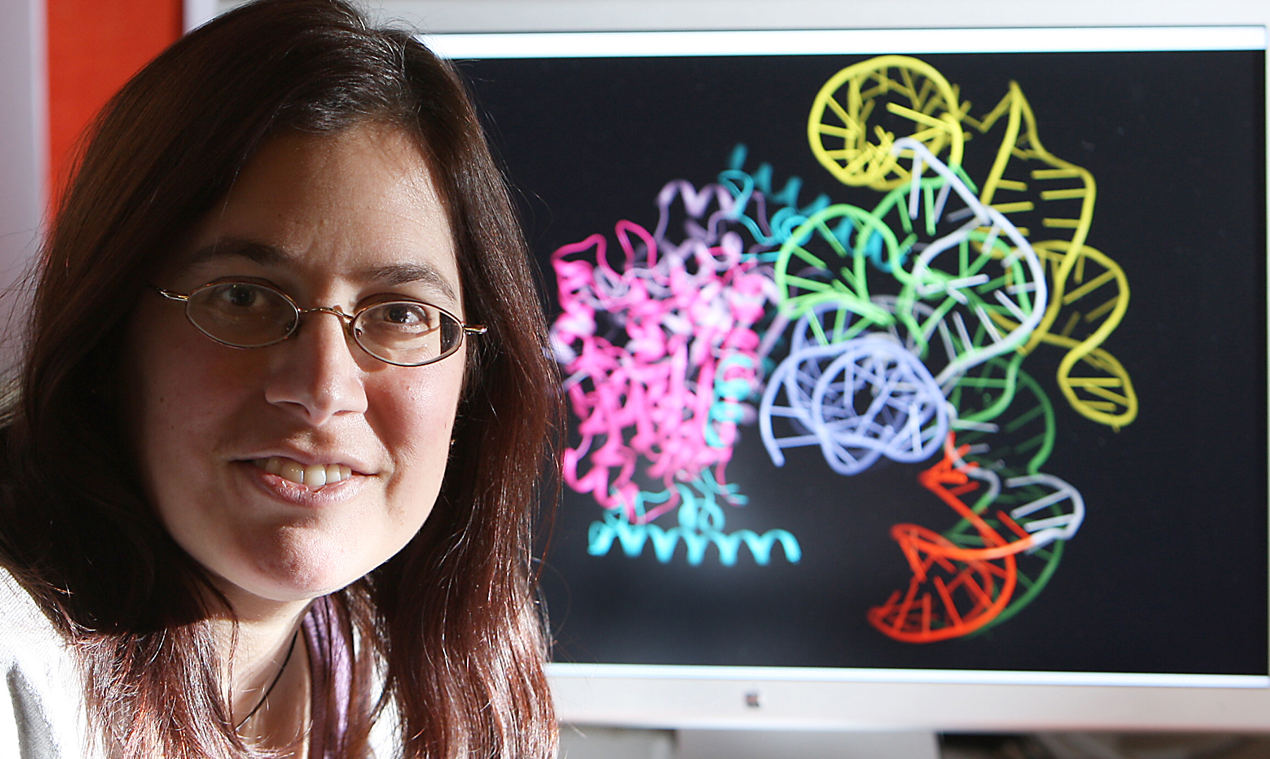
PHOTO CAPTION:
A team led by Purdue structural biologist Barbara Golden crystallized a fungus molecule that has allowed researchers to visualize a stage of evolution. Researchers from Purdue and the University of Texas at Austin collaborated to study the change from a world of RNA to one of RNA and proteins and DNA. (Purdue Agricultural Communication photo/Tom Campbell)
A publication-quality photo is available at https://www.purdue.edu/uns/images/+2007/golden-crystal.jpg
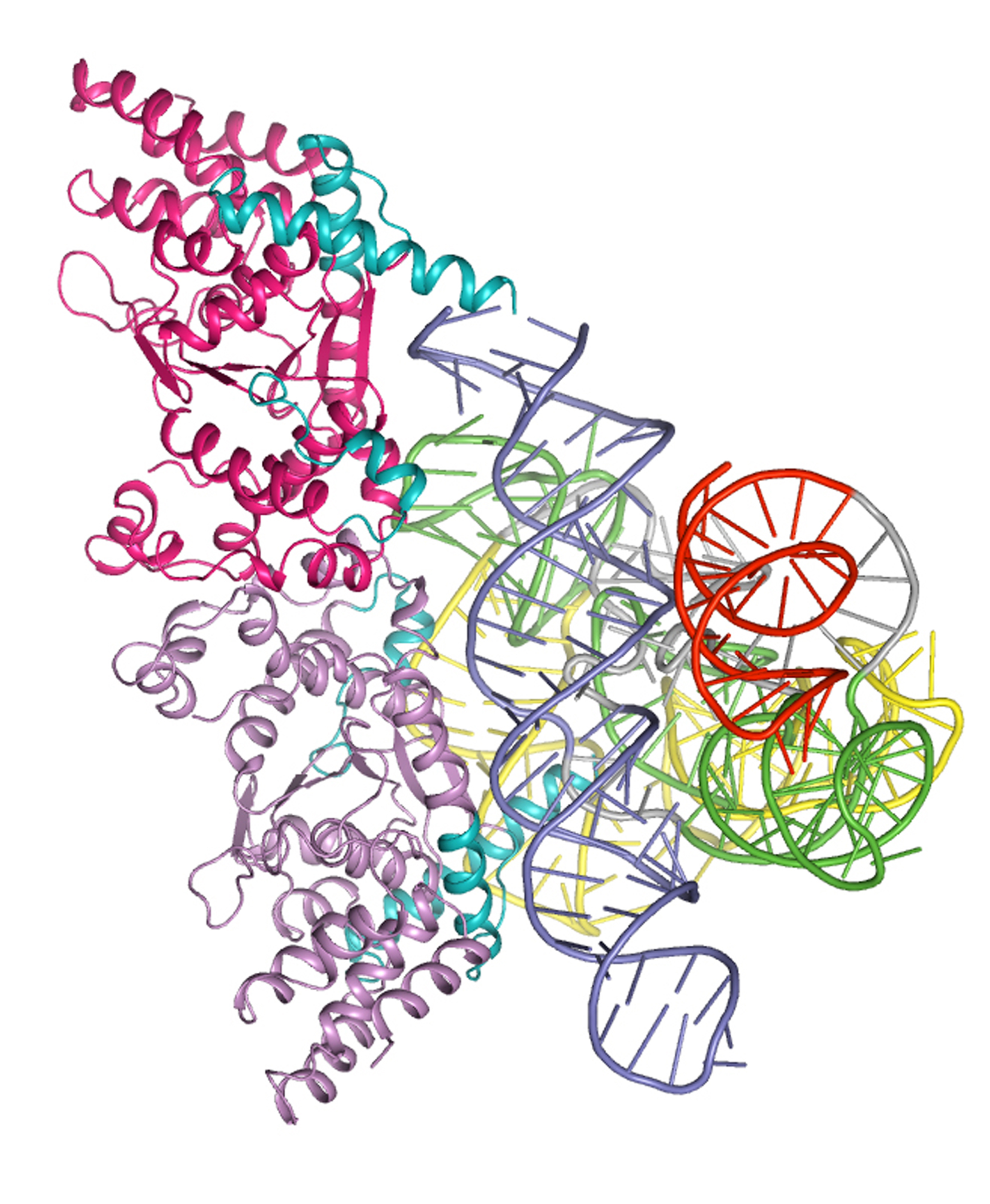
IMAGE CAPTION:
The crystal structure of an RNA molecule bound to a protein was used by Purdue and University of Texas at Austin researchers to study a stage of evolution. (Image courtesy of Barbara Golden, Purdue University Department of Biochemistry)
A publication-quality image is available at https://www.purdue.edu/uns/images/+2007/golden-introncrystal.jpg
Cocrystal Structure of a Tyrosyl-tRNA Synthetase Splicing Factor with a Group I Intron RNA
Paul J. Paukstelis, Jui-Hui Chen, Elaine Chase, Alan M. Lambowitz*, and Barbara L. Golden*
The RNA world hypothesis holds that during evolution structural and functional roles played initially by RNA were assumed by proteins, leading to the latter's domination of biological catalysis. This progression can still be seen in modern biology, where ribozymes, such as the ribosome and RNase P, have evolved into protein-dependent RNA catalysts (RNPzymes). Similarly, group I introns use RNAcatalyzed splicing reactions, but many function as RNPzymes bound to cellular proteins that stabilize their catalytically active RNA structure1, 2. One such protein, the Neurospora crassa mitochondrial tyrosyl-tRNA synthetase (mt TyrRS; CYT-18), is bifunctional and both aminoacylates mt tRNATyr and promotes the splicing of mt group I introns3. Here, we determined a 4.5-Å cocrystal structure of the Twort orf142-I2 group I intron ribozyme bound to splicing-active, C-terminally truncated CYT-18. The structure shows that the group I intron binds across the two subunits of the homodimeric protein using a new RNA-binding surface distinct from that which binds tRNATyr. This RNA binding surface provides an extended scaffold for the phosphodiester backbone of the conserved catalytic core of the intron RNA, allowing the protein to promote the splicing of a wide variety of group I introns. Notably, the group I intron-binding surface includes three small insertions and additional structural adaptations relative to non-splicing bacterial TyrRSs, indicating a multi-step adaptation for splicing function. The cocrystal structure provides insight into how CYT-18 promotes group I intron splicing, how it evolved to have this function, and how proteins could have incrementally replaced RNA structures during the transition from an RNA to RNP world.
To the News Service home page
![]()