
Purdue researchers find 'switch' for skeletal-muscle atrophy
WEST LAFAYETTE, Ind. — Researchers in Purdue University's School of Veterinary Medicine have discovered genetic and drug-treatment methods to arrest the type of muscle atrophy often caused by muscle disuse, as well as aging and diseases such as cancer.
|
"The weight loss and muscle wasting that occurs in patients with cancer or other diseases seriously compromises their well-being and is correlated with a poor chance for recovery," Pond said. "In addition, muscle weakness caused by atrophy during aging can lead to serious falls and bone loss. Exercise is the most beneficial strategy to treat atrophy. However, many individuals are too ill to adequately participate in exercise programs.
"We've found a chemical 'switch' in the body that allows us to turn atrophy on, and, from that, we also have learned how to turn atrophy off."
Findings based on the research, funded in large part by the American Heart Association, are detailed in a study available online today (Wednesday, May 24) in The FASEB Journal, published by the Federation of American Societies for Experimental Biology. The study will be in the journal's print edition in July.
The research team found atrophy of skeletal muscle in mice could be inhibited with both gene therapy and drug treatment using astemizole (as-TEM-uh-zole), an antihistamine. This new insight has potential in many different areas of research, Pond said.
"We have discovered a direct link between atrophy and a protein in the skeletal muscle," Pond said. "This led us to develop methods that would block the protein's ability to cause atrophy. Through drug treatment, we were able to block atrophy, allowing muscle to retain 97 percent of its original fiber size in the face of atrophy."
Astemizole, which was withdrawn from the market in 2000 because of its potential to cause serious cardiovascular problems, wouldn't be suitable for use in humans, Pond said. The drug can be used in mice because it doesn't affect their hearts to the same extent.
"Astemizole administration to humans poses too great a risk," Pond said. "There's a need for more study to avoid those side effects, but the key is that we found a protein capable of sensing muscle disuse and initiating atrophy."
In the drug study, researchers used four groups of mice: a control group, a second group that was given astemizole, and two additional groups in which muscle atrophy was introduced. One of these two groups received astemizole while the second did not. Both of these groups were placed in cages constructed to elevate them so that they were unable to place any weight on their back legs.
"Use of the custom cages to produce atrophy was established in the '80s for simulation of NASA space flight; you can't mimic these effects on muscle and bone in cell culture," said Kevin Hannon, associate professor of developmental anatomy and one of the study's authors. "The mice were able to move around the cage and eat and drink on their own. We monitored their food and water intake and overall health and ensured that they were playing and eating normally."
This method allowed the scientists to demonstrate the effects of skeletal muscle atrophy and investigate reasons for the link with the Merg1a protein. The Merg1a protein is a channel that normally passes a small electrical current across the cell.
The researchers implanted a gene into the skeletal muscle that resulted in a mutant form of this protein that combines with the normal protein and stops the current. The researchers found that the mutant protein would inhibit atrophy in mice whose ability to use their back legs was limited.
Because gene therapy is not yet a practical treatment option in humans, the researchers decided to go a step further and stop the function of the protein with astemizole, which is a known "Merg1a channel blocker." The researchers found that the drug produced basically the same results as the gene therapy. In fact, muscle size increased in mice in the group that were given the drug without any other treatment.
"We are now looking at the differences in the structure of the channel in heart and skeletal muscle to give us clues on how to specifically target muscles without the cardiac side effects," Pond said.
This research also was partially supported by the U.S. Department of Agriculture and Purdue's basic medical sciences department.
Writer: Maggie Morris, (765) 494-2432, maggiemorris@purdue.edu
Sources: Amber Pond, (765) 496-6185, pond@purdue.edu
Kevin Hannon, (765) 494-5949, hannonk@purdue.edu
Purdue News Service: (765) 494-2096; purduenews@purdue.edu
Note to Journalists: An electronic copy of the research paper is available from Maggie Morris, Purdue News Service, (765) 494-2432, maggiemorris@purdue.edu.
PHOTO CAPTION:
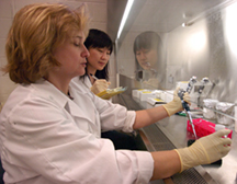
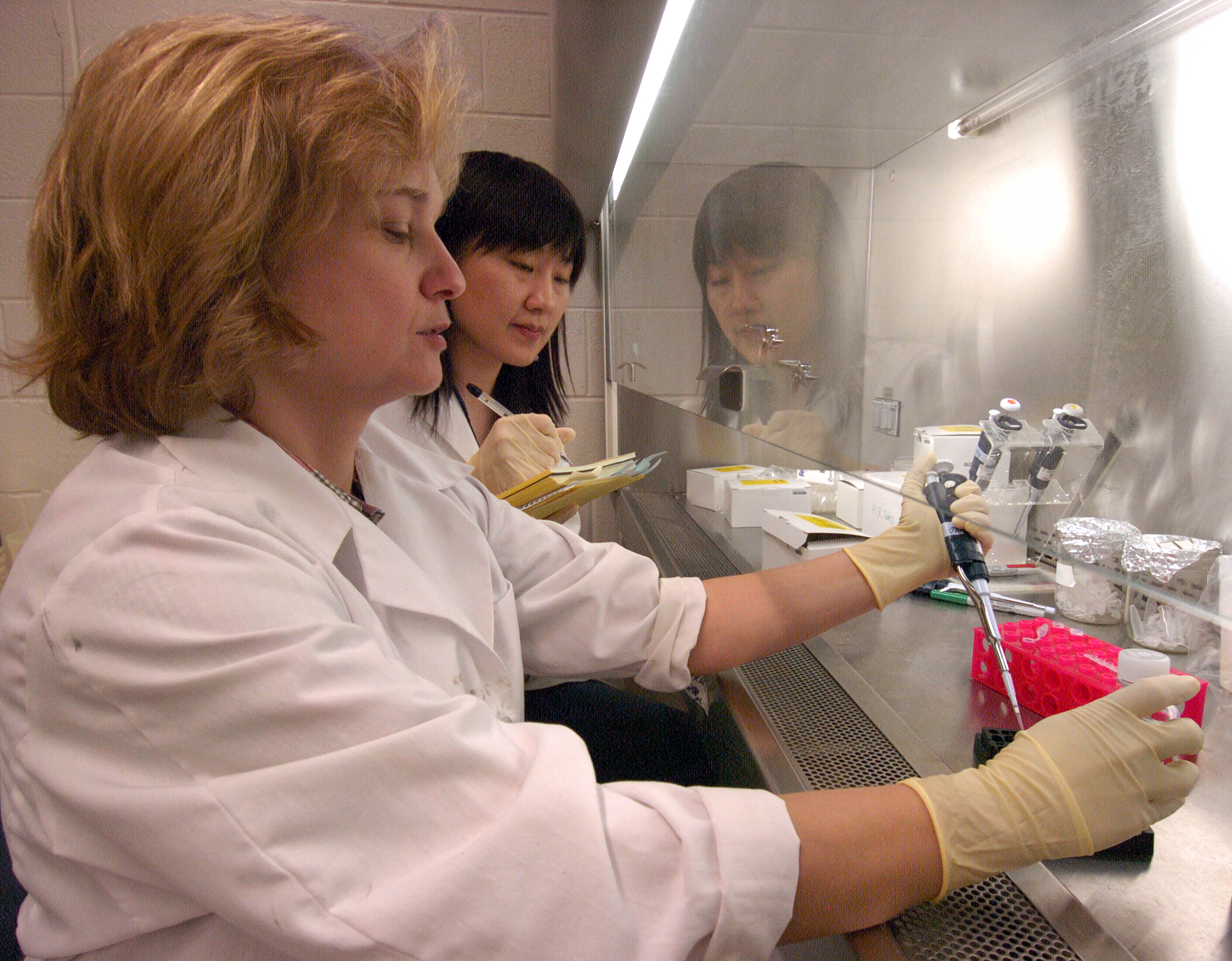
Amber Pond, a research scientist at Purdue University's School of Veterinary Medicine, tests skeletal muscle and heart tissue as Xun Wang, a graduate student in basic medical sciences, takes notes. The two are part of a research team investigating treatments that arrest the muscle atrophy caused by cancer and other diseases. (Purdue News Service photo/David Umberger)
A publication-quality photo is available at https://www.purdue.edu/uns/images/+2006/pond-atrophy.jpg
Merg1a K+ Channel Induces Skeletal Muscle Atrophy
by Activating the Ubiquitin Proteasome Pathway
These authors contributed equally to this work.
Skeletal muscle atrophy results from an imbalance in protein degradation and protein synthesis and occurs in response to injury, various disease states, disuse and normal aging. Current treatments for this debilitating condition are inadequate. More information about mechanisms involved in the onset and progression of muscle atrophy is necessary for development of more effective therapies. Here we show that expression of the mouse ether-a-go-go related gene (Merg1a) K+ channel is up regulated in skeletal muscle of mice experiencing atrophy as a result of both malignant tumor expression and disuse. Further, ectopic expression of Merg1a in vivo induces atrophy in healthy weight bearing mice while expression of a dysfunctional Merg1a mutant suppresses atrophy in hindlimb suspended mice. Treatment of hindlimb suspended mice with astemizole, a known Merg1a channel blocker, inhibits atrophy in these animals. Importantly, in vivo expression of Merg1a in mouse skeletal muscle activates the ubiquitin proteasome pathway that is responsible for the majority of protein degradation that causes muscle atrophy, yet expression of a dysfunctional Merg1a mutant decreases levels of ubiquitinproteasome proteolysis. Thus, expression of Merg1a likely initiates atrophy by activating ubiquitin-proteasome proteolysis. This gene and its product are potential targets for prevention and treatment of muscle atrophy.
To the News Service home page