
July 20, 2005
Machines, software model helping to create better spinal implants
WEST LAFAYETTE, Ind. – Mechanical and biomedical engineers at Purdue University have developed specialized hydraulic machines and software to help industry create better and longer lasting implants for people suffering from spinal injuries, disease and age-related wear.

|
Orthopedic device companies are trying to design implants that behave more like natural disks and tendons in the spine, said Ben Hillberry, a professor of mechanical engineering who is leading the research and working with Purdue's Weldon School of Biomedical Engineering.
The engineers are using machines to test implants for both the lower back, or lumbar region of the spine, and the cervical spine, or the neck region. Implants are attached to spines from cadavers, and then the spines are tested in the Purdue Spine Simulator, a hydraulic machine that recreates the spine's natural movements and shows how the implants stand up to everyday activities.
Data from the experiments also are being used to validate a complex computer model, which companies will use to test implants. The software is a "finite element model," an application widely used in industry that contains a series of geometric shapes, such as rectangles and triangles, each providing specific data describing a part's strength and other characteristics.
"Creating implants for the spine presents interesting challenges, different from those encountered in implants for other parts of the body, such as the hips and knees," said Eric Nauman, an assistant professor of mechanical engineering.
Some implants made of titanium and other materials are used to treat conditions such as arthritis, in cases when a portion of the spine has been removed. Bracket-like implants called "pedicle bridges" help to keep the spine stable after the diseased portion has been removed. Other implants include artificial disks to replace disks that are damaged from wear or disease in the lower back and cervical region.
One of the machines designed by the Purdue engineers is used specifically to test implants for the cervical spine.
"There is much more movement in the cervical spine than in the lumbar portion, so what we are primarily testing with this machine is how well implants will stand up to wear over a period of about 10 years," said Shreekant Gayakar, a graduate student in mechanical engineering. "We are replicating the range of motions seen in the human neck."
The machine tests ball-and-socket-like implants such as the ones inserted during surgeries to replace damaged parts in the cervical spine.
"In order for implants to be approved by the Food and Drug Administration, it has to be shown that they can last 10 million cycles, or 10 million movements, which translates into about 10 years of living," Hillberry said. "Our goal is to complete 10 million cycles over a four-month period."
The Purdue researchers are putting the machines through their paces to meet standards set by the American Society for Testing and Materials.
The cervical spine machines are designed so that many machines could be linked together in a series, enabling companies to test different implants under the same conditions and duration.
Whereas wear is the primary concern in the cervical spine, damage from injury and disease are the biggest problems in the lower back.
The spine simulator machine recreates the stresses and range of motions encountered by the human spine, including twisting and bending in various directions, said Jeremie Wade, a graduate student in mechanical engineering who is involved in research using the machine. The simulator was inspired by orthopedic surgeon John Gorup, who practices in Lafayette, Ind., and was designed by Beth Galle, who graduated from Purdue with a master's degree in mechanical engineering in 2004.
"Not many machines can duplicate the motions of the human spine – I know of only one on the market," Wade said.
Researchers also are using the spine simulator to test implants that replace diseased "facet joints." The facet joints are often damaged by arthritis and usually are treated by injecting them with compounds to relieve the pain. Unlike other arthritic joints, such as hips, knees and shoulders, facet joints have not, until recently, been removed and replaced with artificial implants.
"Arthritis in your spine is awful," Nauman said. "It is one of the worst kinds of skeletal degeneration you can get."
Current treatments for some spinal conditions involve fusing several vertebrae together.
"The problem with fused vertebrae is that because you fuse parts together and things don't move the way they used to, the surrounding vertebrae eventually become damaged, and you sort of set yourself up for more medical complications and procedures in the future," Nauman said. "What doesn't exist right now is a really nice way to restore the natural motion of the spine, relieve the pain and prevent damage to surrounding portions of the spine.
"Basically, the medical community is trying to create new devices that duplicate the spine's natural motion instead of immobilizing the damaged area."
Hillberry and Kimberly Campana, a biomedical engineering doctoral student, are working with Archus Orthopedics Inc., a company in Redmond, Wash., to develop mathematical simulation tools that will allow the accelerated development of next-generation implants.
"Future designs may ultimately be used in conjunction with total disk replacements, allowing the restoration of function at any vertebral level in the spine, much like total hip and total knee replacement currently achieve, "said Jorge Ochoa, vice president of research and development and chief technology officer at Archus Orthopedics.
To that end, the company recently has obtained approval to begin implanting its Total Facet Arthroplasty System in Europe and will begin an FDA-approved clinical trial of the device in the United States this summer.
The system is designed to allow doctors to manipulate the lumbar spine surgically to reduce pressure on the nerves by removing any tissue that may compress them and by eliminating other sources of pain, including arthritic facets, without having to fuse the spine to regain stability, Ochoa said.
"The application of three-dimensional computational models, such as the one being developed at Purdue, will enable companies like Archus to reduce the cost of developing implants and increase the speed of new product realization," Ochoa said.
Doctors currently are using prosthetic disks, but it is difficult to insert them into the spine.
"These implants are tricky to insert because major arteries are located along the spine," Hillberry said. "If an implant is not designed to fit precisely into a particular person's spine, the spine can actually eject it during surgery, causing the artificial part to sever a major blood vessel that runs along the spine.
"If that happens, the patient could die on the operating table. One benefit the model provides is that it can tell you if the implant is going to be ejected before it is put in patients."
The 3-D model also promises to help reduce costs for companies.
"A company designing an implant can test it in this model," Campana said. "It's fairly fast and inexpensive to use, so companies can get an idea of how their implant will perform, and they can make small modifications without having to order 10 cadavers, which are costly.
"Companies can use this model as a virtual test machine."
Relying on tests with cadaver spines has a major limitation: the spines lack the stability of muscles that exist in real-life spines, which contain a complex network of vertebrae, ligaments and muscles. The Purdue researchers are now adding muscles to their spine simulation model to make it more realistic.
"This is extremely difficult because you are trying to model the interactions of all the ligaments, muscles, cartilage and bones in the spine," Nauman said.
The model also will provide a new tool for researchers studying osteoporosis.
"The one limitation that the spine simulator has is that it can't be used to test spines afflicted with osteoporosis because they are too fragile," Nauman said. "So Kim is going to be able to model what happens in a spine with osteoporosis.
"With the model, we can study the performance of implants used to treat people with osteoporosis, and we can also test the effects of certain drug therapies that are supposed to add bone to the spine."
The model also can be used to help develop better ways to straighten the spine for people who have scoliosis, or a curvature of the spine.
Hillberry began research to develop artificial knees during the 1970s and started developing the computational spine model in 1992. The model delves into "kinematics," or the precise motions of the spine.
Earlier work to develop the computational model was funded through the Indiana 21st Century Research and Technology Fund, established by the state of Indiana to promote high-tech research and to help commercialize innovations.
The engineers are working in ongoing research with the Center for Paralysis Research in Purdue's School of Veterinary Medicine.
Writer: Emil Venere, (765) 494-4709, venere@purdue.edu
Sources: Ben Hillberry, (765) 494-5721, (765) 714-6297 (cell), hillberr@ecn.purdue.edu
Eric Nauman, (765) 494-8602, enauman@purdue.edu
Jorge Ochoa, (425) 869-2100, ext. 246, jochoa@archususa.com
Kim Campana, kcampana@purdue.edu
Jeremie Wade, jawade@purdue.edu
Shreekant Gayakar, sgayaka@purdue.edu
Purdue News Service: (765) 494-2096; purduenews@purdue.edu
Related Web site:
Purdue University Home Page
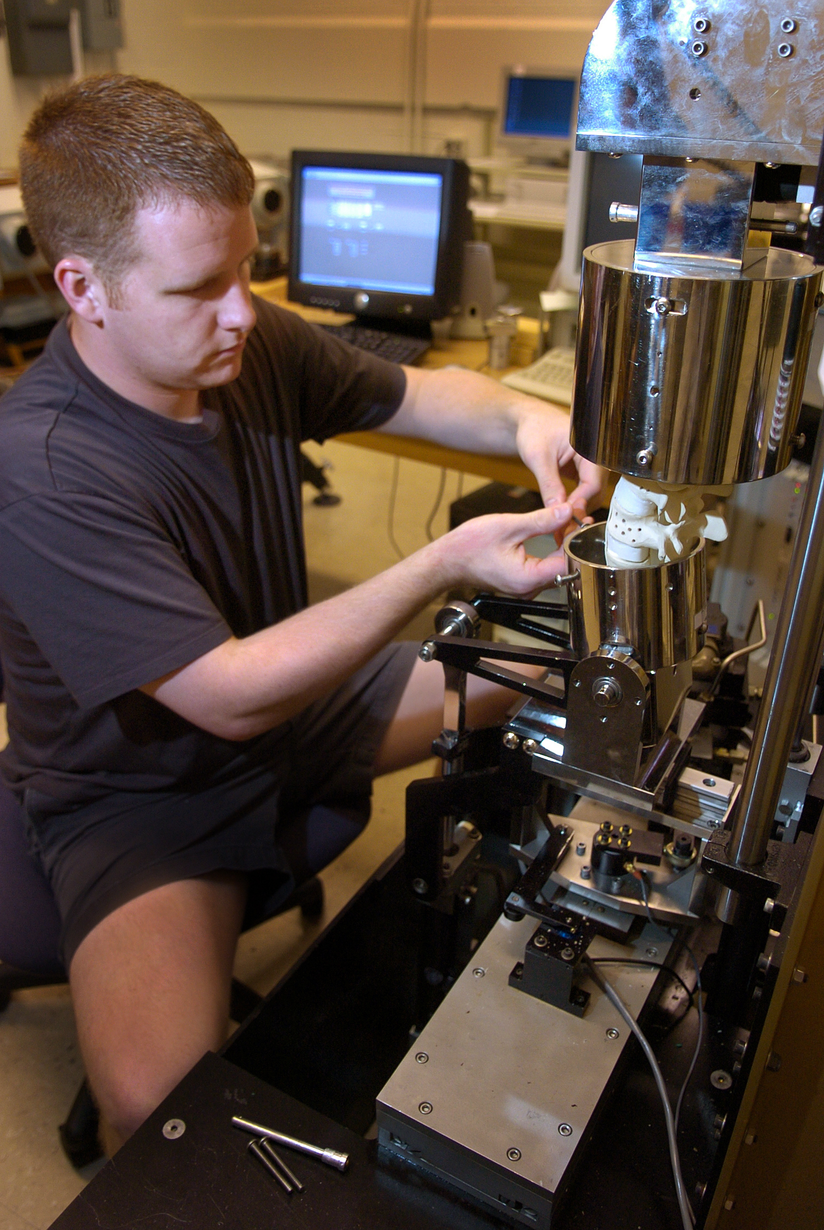
PHOTO CAPTION:
Jeremie Wade, a graduate student in mechanical engineering at Purdue University, makes adjustments to the Purdue Spine Simulator, a hydraulic machine that recreates the spine's natural movements and shows how spinal implants stand up to everyday activities. Mechanical and biomedical engineers have developed specialized hydraulic machines and software to help industry create better and longer lasting implants for people suffering from spinal injuries, disease and age-related wear.
(Purdue News Service photo/David Umberger)
A publication-quality photo is available at https://www.purdue.edu/uns/images/+2005/hillberry-nauman-spine.jpg
To the News Service home page