 Purdue News
Purdue News|
|
|
April 9, 2004 Self-assembling 'nanotubes' offer promise for future artificial jointsWEST LAFAYETTE, Ind. – Tiny "nanotubes" that assemble themselves using the same chemistry as DNA could be ideal for creating better artificial joints and other body implants.
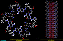
Researchers at Purdue University, the University of Alberta and Canada's National Institute for Nanotechnology have discovered that bone cells called osteoblasts attach better to nanotube-coated titanium than they do to conventional titanium used to make artificial joints. "We have demonstrated the same improved bone-cell adhesion with other materials, but these nanotubes are especially promising for biomedical applications because we'll probably be able to tailor them for specific parts of the body," said Thomas Webster, an assistant professor of biomedical engineering at Purdue. Findings are detailed in a paper appearing in the April issue of Nanotechnology, published by the Institute of Physics in the United Kingdom. The paper was written by Purdue biomedical engineering doctoral student Ai Lin Chun, Purdue chemistry doctoral student Jesus G. Moralez, Webster and Hicham Fenniri, a professor of chemistry at the University of Alberta and senior research officer at the Canadian Nanotechnology institute, where Chun and Moralez are doctoral students as well. The self-assembling nanotubes were developed by Fenniri while he was an assistant professor at Purdue. Webster has shown in a series of experiments that bone cells and cells from other parts of the body attach better to various materials that possess surface bumps about as wide as 100 nanometers, or billionths of a meter. Conventional titanium used in artificial joints has surface features on the scale of microns, or millionths of a meter, causing the body to recognize them as foreign and prompting a rejection response. The body's rejection response eventually weakens the attachment of the implants and causes them to become loose and painful, requiring replacement surgery. The nanometer-scale bumps mimic surface features of proteins and natural tissues, not only prompting cells to stick better but promoting the growth of new cells. Bone and other tissues adhere to artificial body parts by growing new cells that attach to the implants, so the experiments offer hope in developing longer lasting and more natural implants, Webster said. Now researchers have discovered that the self-assembling nanotubes represent an entirely new and potentially superior material to use for artificial body parts. Fenniri created the self-assembling structures by using the chemistry of deoxyribonucleic acid, or DNA, to make a series of molecules that are "programmed" to link in groups of six to form tiny rosette-shaped rings. Numerous rings then combine to create the rod-like nanotubes, which have widths of only about 3.5 nanometers. "He had these nice nanotubes, and I had this work that showed nice bone synthesis and other tissue regeneration on nanomaterials, so we said, 'Wouldn't it be great to actually combine the two to see if his material can promote new bone growth with these nanotubes?'" Webster said. One nanometer is roughly the length of 10 hydrogen atoms strung together. A human hair is more than 30,000 times wider than the rosette nanotubes used in the study. Self-assembly is a well-known principle in biology in which the right mix of molecules interact on their own to form distinctive structures ranging from DNA to cells and organs. The rosette-shaped rings are made of guanine and cytosine, which are molecules called "base pairs" that come together to form DNA. In addition to possible biomedical applications, the nanotubes offer promise in the design of future materials, electronic devices and drug delivery systems. The researchers coated titanium with the nanotubes and placed them in Petri plates containing a liquid suspension of bone cells colored with a fluorescent dye. After a few hours, the nanotube-coated titanium was washed, and a microscope was used to count how many of the dyed osteoblasts adhered to the material. Out of 2,500 bone cells in the suspension, 2,300 to 2,400 were found to adhere to the nanotube-coated metal. That compares with about 1,500 cells adhering to titanium not coated with the nanotubes, representing an increase of about one-third. The quick attachment of bone cells is critical to create a solid bond between orthopedic implants and the body's natural bone. The same applies to artificial parts transplanted in other parts of the body, such as arteries and the brain. "The reason we are so excited is that we see improved osteoblast function on the coated titanium compared to the plain titanium," Webster said. Webster has found similar results with other materials that possess the nano-scale surface bumps, such as ceramics, metals and nanotubes made of carbon. The rosette nanotubes, however, may provide a major advantage over those materials, he said. Protein components, such as "signaling peptides," or amino acids, such as lysine and arginine, can be easily attached to the surface of the nanotubes, making it feasible to tailor the nanotubes so that they are recognized by specific cells and body parts. "There are definite amino acid sequences that bone cells recognize and stick to," Webster said. "One of those sequences is arginine, glycine and aspartic acid. There is a lot of work in the field now to incorporate this sequence into materials. "One of the other reasons we were so excited about this is that we can put this sequence on these tubes." Attaching the sequence of amino acids onto the nanotubes will likely increase osteoblast adhesion even more, Webster said. Various parts of the body recognize and attach to different sequences. "I think this really points to strong biomedical applications," Webster said. "If the cells you are targeting respond to protein sequence XYZ, you just put that sequence on the nanotubes and you can promote this attachment." Another finding in the research is that low concentrations of the nanotubes provide the same results as higher concentrations. "That means you can use very low concentrations of this and still get statistically higher bone-cell attachment," Webster said. "So it's cheap. You don't need a lot of it to get the effect that you want." Unlike other nano-scale materials Webster has worked with, the rosette nanotubes automatically arrange themselves into a webbed pattern on the surface of the titanium. The pattern resembles those seen by natural collagen fibers in bones and other tissues. Future work will focus on further modifying the nanotubes and conducting additional experiments. The need for better technology is growing as more artificial body parts are used, Webster said. For example, about 152,000 hip replacement surgeries were performed in the United States in 2000, representing a 33 percent increase from 1990. The number of hip replacements by 2030 is expected to grow to 272,000 in this country alone because of aging baby boomers. The research has been funded by the National Science Foundation, American Chemical Society, Purdue Research Foundation, Whitaker Foundation, 3M Co. and Canada's National Institute for Nanotechology. Writer: Emil Venere, (765) 494-4709, venere@purdue.edu Sources: Thomas Webster, (765) 496-7516, twebster@purdue.edu Hicham Fenniri, (780) 492-8988, hicham.fenniri@ualberta.ca Purdue News Service: (765) 494-2096; purduenews@purdue.edu Note to Journalists: An electronic or hard copy of the research paper is available from Emil Venere, (765) 494-4709, venere@purdue.edu. The paper also can be downloaded at the Nanotechnology magazine Web site.
Previous releases about Thomas Webster's work: Purdue research suggests 'nanotubes' could make better brain probes Designer molecules link together to make nanotubes a snap
IMAGE CAPTION: A publication-quality photo is available at https://ftp.purdue.edu/pub/uns/+2004/webster-rosette.jpg Helical rosette nanotubes: a more effective orthopedic implant material and Thomas J. Webster1 1 Department of Biomedical Engineering, Purdue University 2 National Institute for Nanotechnology and Department of Chemistry, National Research Council and the University of Alberta Due to the nanometric properties of some physiological components of bone, nanomaterials have been proposed as the next generation of improved orthopedic implant materials. Yet current efforts in the design of orthopedic materials such as titanium (Ti) are not aimed at tailoring their nanoscale features, which is now believed to be one reason why Ti sometimes fails clinically as a bone implant material. Much effort is thus being dedicated to developing improved bioactive nanometric surfaces and nanomaterials for biospecificity. Helical rosette nanotubes (HRN) are a new class of self-assembling organic nanotubes possessing biologically-inspired nanoscale dimensions. Because of their chemical and structural similarity with naturally-occurring nanostructured constituent components in bone, such as collagen and hydroxyapatite, we anticipated that an HRN-coated surface may simulate an environment that bone cells are accustomed to interacting with. The objective of the present in vitro study is therefore to determine the efficacy of Hrn as a bone prosthetic material. Results of this study clearly show that both HRN-K1 and HRN-Arg coated Ti displayed enhanced cell adhesion when compared to uncoated Ti. Enhanced cell adhesion was observed even at concentrations as low as 0.005 mg ml-1. These results point towards new possibilities in bone tissue engineering as they serve as a starting point for further mechanistic studies as well as future manipulating of the outer chemistries of HRN to improve the results beyond those presented here. One such effort is the incorporation of peptide sequences on the outer surfaces of HRN and/or growth factors known to enhance bone functions.
|
