 Purdue News
 Purdue News
 Cathrin De Nooyer, a master's student in Purdue's Department of Earth and Atmospheric
Sciences, has developed a methodology that takes the guesswork out of using computerized
axial tomography -- a system designed to analyze soft tissues and bone -- to obtain three-dimensional images of the fossils contained in chunks of various types of rock.
Cathrin De Nooyer, a master's student in Purdue's Department of Earth and Atmospheric
Sciences, has developed a methodology that takes the guesswork out of using computerized
axial tomography -- a system designed to analyze soft tissues and bone -- to obtain three-dimensional images of the fossils contained in chunks of various types of rock.
"CAT scans are expensive, and it's hard to get access to the equipment, but if you can maximize your time spent, you can get some really wonderful pictures," she says.
De Nooyer, from Sacramento, Calif., has traveled to hospitals throughout Indiana to use a variety of different scanners to assist in her studies of small invertebrate fossils. Working with an interdisciplinary team at Purdue, she has established a set of procedures that will allow almost any make or model of scanning machine to penetrate various types of rock on almost any fossil specimen.
"My goal was to establish a protocol so that any interested paleontologists could go to their local hospital and request the right procedures to get the best images of their particular fossil type," she says.
She presented information on her methodology Oct. 30 at the annual meeting of the Geological Society of America in Denver.
CAT scans can produce images of various tissues in the body by passing radiation through the body and measuring the amount of radiation absorbed by the tissue. Because tissues vary in density -- for example, bone is much denser than muscle -- they will absorb different amounts of radiation. A computer then converts the data into a three-dimensional picture.
The same principles can be applied to fossils, which are made up of rock and petrified bone or tissue. "For example, we can detect a skull within a rock because it is made of different materials than the rock," De Nooyer says.
Generally, CAT scanners are calibrated for scans of humans. Through a process of trial-and-error, De Nooyer has established a series of algorithms and settings to obtain the best images of fossils. She also has learned how to "read" the images to interpret the shadows and shadings that appear.
"We're learning to be blob-ologists," she says. "Doctors have had the luxury of looking at human bodies for a long time, so anatomically speaking, they know where things are located. When looking at a fossil, we know what we hope to be on the inside, but it may not be there. So we're learning to critically evaluate the various shadows and images to get a clear view of what's inside."
The method allows scientists to view the internal structure of a fossil without risking damage to the specimen.
"Normal protocol would be to spend hours and hours chiseling rock away from a specimen, running the risk of destroying it in the process," De Nooyer says. "Another technique is to make serial sections, by slicing off thin layers of the fossil and putting them on slides to visually analyze. This method always results in a permanently destroyed specimen."
A CAT scanner also allows scientists to analyze more specimens in a short amount of time.
"It would take a single person several days to section a fossil mollusk into thin sections," De Nooyer says. "After that, you would still have to read the sections and trace them to get a picture of the fossil. Using the most sophisticated medical CAT scanner available, I can have clear images in four seconds."
While CAT scans have seen limited use in studies on fossils and mummies, De Nooyer is the first to adapt the method to examine invertebrates. She studies ancient mollusks, clams and snails that have been collected from Antarctica, Europe and Indiana. By analyzing these samples, she can infer what the climate was like when the creatures lived hundreds of millions of years ago.
In addition, she has analyzed a dinosaur egg and the skull of a small, fox-like mammal.

"It's exciting. You can go in there with a rock specimen and come out a few minutes later with a perfect three-dimensional view of what's inside the rock," she says.
CAT scans first were used in paleo-anthropologic studies the late 1970s to obtain images of mummies. During the past decade, scientists have made various attempts to apply the technology to fossils embedded in rock, but efforts have been hindered by problems such as lack of power to penetrate the rock.
"Five years ago, medical scanners just didn't have enough juice," De Nooyer says. "With the newer machines, we're able to penetrate the rock much better. Also, scan times have become incredibly fast; what used to take six minutes now takes 60 seconds."
To fine-tune her method, De Nooyer consulted with experts from various fields, including computer sciences, mineralogy, and medicine.
Source: Cathrin De Nooyer, (765) 494-3704; e-mail, denooyer@alka.cs.purdue.edu
Writer: Susan Gaidos, (765) 494-2081; e-mail, susan_gaidos@purdue.edu
Purdue News Service: (765) 494-2096; e-mail, purduenews@purdue.edu
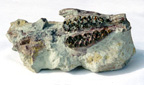
NOTE TO JOURNALISTS: Three color photographs are available. The first shows Cathrin De Nooyer at a workstation, download here, stacking "slices" of data from a CAT scan to construct a three-dimensional image of an invertebrate fossil. The second shows part of a fossilized mammal skull jutting out of a rock, download here, and the third shows a three-dimensional view of the skull, download here.
Photo captions:
Cathrin De Nooyer, a master's student in earth sciences at Purdue, uses computer software
to create a three-dimensional picture of a fossil from CAT scan images. The CAT scan
provides a series of views of thin sections of the sample. The computer then stacks those views like slices of bread in a loaf to generate a full view of the specimen.
(Purdue News Service Photo by David Umberger)
Color photo, electronic transmission, and Web and ftp download available. Photo IDs:
DeNooyer/Cathrin.
Part of a fossilized mammal skull juts out of a rock (above). The remainder of the
skull is buried within the rock. Using CAT scans and computer technology, De Nooyer
was able to construct a series of three-dimensional views of the entire skull (below).
The skull belonged to a small fox-like mammal that lived 35 million to 55 million years
ago.
Color photos, electronic transmission, and Web and ftp download available. Photo IDs:
DeNooyer/Skullscan, DeNooyer/Skullphoto.
