
November 23, 2004
Purdue researchers align nanotubes to improve artificial joints
WEST LAFAYETTE, Ind. – Researchers at Purdue University have shown that artificial joints might be improved by making the implants out of tiny carbon tubes and filaments that are all aligned in the same direction, mimicking the alignment of collagen fibers and natural ceramic crystals in real bones.
|
The researchers already have shown in a series of experiments that bone cells in Petri dishes attach better to materials that possess smaller surface bumps than are found on conventional materials used to make artificial joints. The smaller features also stimulate the growth of more new bone tissue, which is critical for the proper attachment of artificial joints once they are implanted.
Now, the Purdue researchers have shown even more enhanced cell adhesion and growth when so-called "nanotubes" and nanofibers are aligned in the same direction. This orientation is similar to the way collagen and natural ceramic crystals, called hydroxyapatite, are aligned in bone, said Thomas Webster, an assistant professor of biomedical engineering at Purdue.
Findings were presented at two recent scientific conferences in research papers written by Webster; Purdue physics doctoral student Dongwoo Khang; and three researchers from the Seoul National University in South Korea, physics doctoral students Minbaek Lee and Sun Namkung, and physics professor Seunghun Hong.
Previous experiments in the Purdue lab have shown that about one-third more bone-forming cells, or osteoblasts, attach to carbon nanotubes that possess surface bumps about as wide as 100 nanometers, or billionths of a meter. Fewer bone cells stick to conventional titanium, which has surface features on the scale of microns, or millionths of a meter.
The nanometer-scale bumps mimic surface features of proteins and natural tissues, prompting cells to stick better and promoting the growth of new cells, Webster said. The findings also suggest that using such nanometer-scale materials might cause less of a rejection response from the body. Rejection eventually weakens the attachment of implants and causes them to become loose and painful, requiring replacement surgery.
Aligning the nanotubes to further mimic natural bone also might provide more strength, Webster said.
Researchers used two methods to align the tiny nanotube structures, which have diameters of about 60 nanometers. One nanometer is roughly the length of 10 hydrogen atoms strung together. A human hair is more than 1,000 times wider than the nanotubes used in the study.
In one method, researchers mixed the nanotubes in a polymer, or plastic, and passed an electric current through the mixture. Because nanotubes have the same natural electrical charge, they react to electricity by orienting themselves in the same direction. Once the polymer solidifies, the nanotubes are fixed in the aligned position.
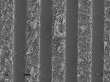
The research team also aligned the nanotubes using another method in which the nanotubes are poured into grids of tiny channels. Because the channels are so narrow, the tubes can only fit lengthwise, causing them to become aligned. The grids can then be removed, leaving behind the aligned nanotubes.
The researchers then added the aligned nanotubes to a suspension of dyed bone cells in a small container. After one hour, the nanotubes were washed and a microscope was used to count how many of the dyed osteoblasts adhered to the material. Out of 3,000 bone cells per square centimeter of surface area, about 80 percent specifically stuck to and aligned with the carbon nanotubes – or about twice as many as those that adhered to non-aligned nanotubes in previous work.
"So, in a very short period of time, one hour, we're already seeing a big improvement in how well the cells stick to the nanotubes," Webster said.
Future research may focus on combining the two methods for aligning nanotubes. Using the grid technique creates a greater number of aligned nanotubes on the surface, which helps to increase bone-cell adhesion and alignment, whereas using electricity could better stimulate the growth of new bone tissue.
The research has been funded by the National Science Foundation though the NSF Nanoscale Exploratory Research program.
Findings were presented in October during the Biomedical Engineering Society's annual meeting and a conference by the Society for Biomaterials entitled Biomaterials in Regenerative Medicine: The Advent of Combination Products. Both meetings were in Philadelphia.
Writer: Emil Venere, (765) 494-4709, venere@purdue.edu
Source: Thomas Webster, (765) 496-7516, twebster@purdue.edu
Purdue News Service: (765) 494-2096; purduenews@purdue.edu
Note to Journalists: An electronic or hard copy of the research paper is available from Emil Venere, (765) 494-4709, venere@purdue.edu.
Related Web site:
Nano Letters
IMAGE CAPTION:
This image, taken with a scanning electron microscope, shows arrays of nanofibers that have been aligned by pouring them into grids of tiny channels. Because the channels are so narrow, the tubes can only fit lengthwise, causing them to become aligned, similar to the way in which collagen fibers and natural ceramic crystals are aligned in bones. Because the nanofibers are aligned like natural collagen, they might be used to create better artificial joints that last longer and attach better to human bones. (Purdue University, Department of Biomedical Engineering and Department of Physics)
A publication-quality photograph is available at https://ftp.purdue.edu/pub/uns/+2004/webster-align.jpg
Osteoblasts Aligned with Carbon Nanotubes/Nanofibers
In this work, we describe the first successful alignment of osteoblasts (bone-forming cells) on aligned carbon nanofiber/nanotube arrays in either a polymer matrix or SiO2 substrate. To align carbon nanofibers (CNFs) on a polymer matrix, we developed a novel imprinting method. In addition, the surface-templated assembly method was used to prepare aligned single-wall carbon nanotube (swCNT) arrays on a SiO2 substrate. On both substrates, results demonstrated that aligned carbon nanofiber/nanotube arrays can be used to direct osteoblast morphology important for the continued development of a novel biocompatible substrate with potential for regenerating bone.
To the News Service home page