 Purdue News
Purdue News|
|
August 20, 2004 Virus known for its photo ops makes its movie screen debutWEST LAFAYETTE, Ind. – High-resolution snapshots of a virus attacking its host – which have culminated in a movie of the process – could reveal secrets of viral infection and improve gene therapy techniques, according to a Purdue University research group.
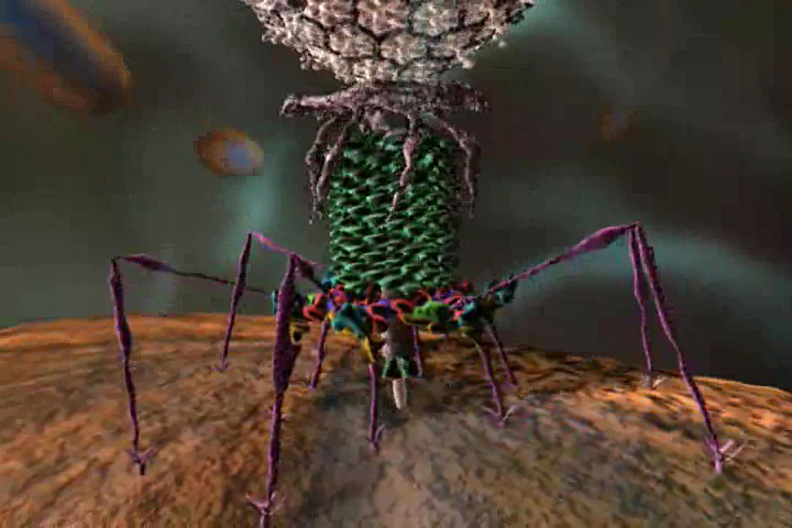
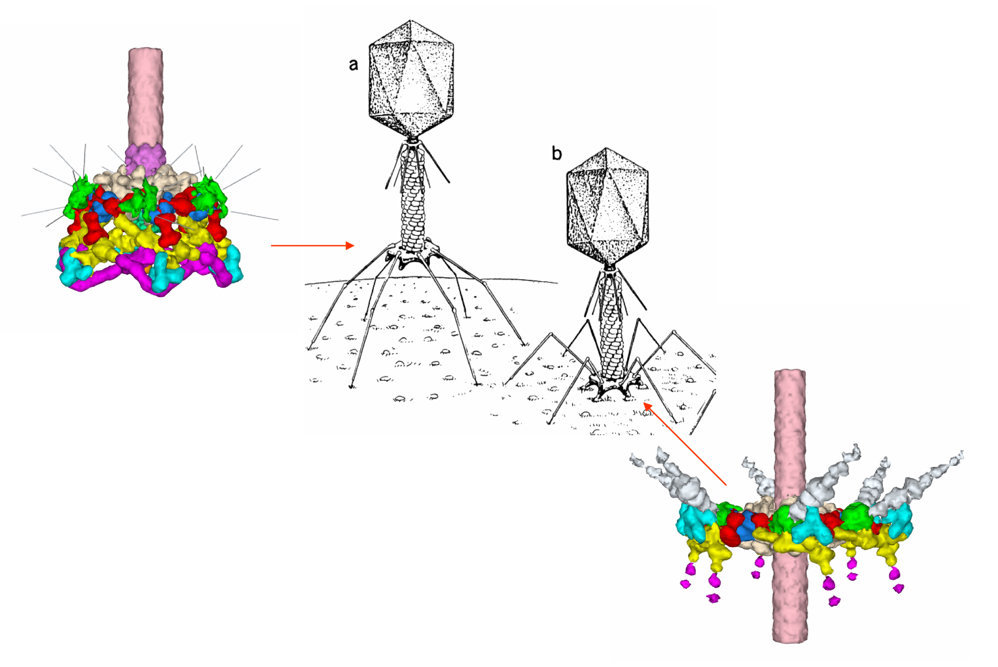
Structural biologists including Michael G. Rossmann have obtained clearer pictures of how the T4 virus, long known to infect E. coli bacteria, alters its shape as it prepares to pierce its host's cell membrane. The complicated infection process requires a flower-like section of the virus, known as the baseplate, to shape-shift by dramatically changing the configuration of the numerous proteins that form it. The team has taken cryoelectron microscope images of the baseplate from different moments in the process and transformed them into a brief animated movie, helping scientists understand how infection occurs and possibly enabling them to apply this knowledge for the benefit of human patients in the future.
"Instead of a still photo of the baseplate, we now have a movie of it opening," said Rossmann, who is Henley Distinguished Professor of Biological Sciences in Purdue's School of Science. "A better understanding of the infection process is a step forward for fundamental science, but it also could allow scientists to alter the baseplate so that the virus could infect cells other than E. coli. T4 might then be used to deliver beneficial genes to damaged or infected human tissue." The research was performed at Purdue and the Institute of Bioorganic Chemistry in Moscow by a team of scientists including first author Petr G. Leiman, Paul R. Chipman, Victor A. Kostyuchenko, Vadim V. Mesyanzhinov and Rossmann. The paper appears in the current (Aug. 20) issue of the scientific journal Cell, and it builds on research the team published last year regarding the baseplate of the T4 virus. This previous paper offered a close-up picture of the baseplate at a single moment in time, information that was valuable because of the detail it provided of the part of the virus that attached itself to E. coli's surface. "It was good to see the baseplate at such unprecedented resolution, but the infection process is not a still picture – it's a story," Rossmann said. "We knew we needed to see more than one scene in that story if we were ever to understand its full meaning." The baseplate is composed of 16 types of protein molecules, most present in multiple copies. Before infection, these proteins fit together to form a hexagonal shape. Together with the 12 legs that extend from the T4's tail to grasp the victim E. coli, the virus resembles an Apollo moon lander. When the T4 approaches "touchdown" on an E. coli's cell membrane, the baseplate's proteins unfold in a complex motion, opening like a flower's petals and changing shape from a hexagon to a star. "We can now visualize how these proteins move together, which means a great deal for anyone trying to comprehend infection," Rossmann said. "If you saw a car speed past you for the first time ever, it might impress you, but you probably wouldn't have much of an idea how it works. But if you stop it, you can examine the engine and find out. That's essentially what we've done – stop the virus at two points in its attack on E. coli and examine the difference." Scientists speculate that viruses are a key player in the evolutionary process on planet Earth. Far from being mere purveyors of disease, the viral infection process also could be partly responsible for spreading new genes among organisms rapidly and preparing their hosts for future environmental changes. This is part of viruses' fascination for scientists like those on Rossmann's team and why some medical professionals seek to use altered viruses to cure illnesses rather than cause them. "Viruses' great talent – injecting genetic material into living cells – could make them valuable for delivering healthy DNA to cells damaged by injury or cancer," said Leiman, a postdoctoral researcher in Rossmann's lab. "T4's baseplate proteins could be altered so it could infect human cells instead of E. coli. This study could bring us one step closer to using it as a gene therapy vehicle." Gene therapy using T4 remains a distant possibility, however, and Rossmann said the true value of the team's latest research was for the fundamental understanding it provides of the viral world. "Viruses are among the tiniest of biological entities, yet nature has designed them to perform very complicated tasks," he said. "Understanding their behavior will open doors for scientists in many disciplines, especially with biologists, chemists and physicists increasingly working side by side." As a step in that direction, Rossmann said he hopes that he and his colleagues will be able to obtain a better picture of the components within the tiny mechanism that is the baseplate. "Our knowledge of the orientation of the proteins within the baseplate could still stand some improvement," he said. "We'd like to look under the hood of this car, so to speak, and determine precisely how the carburetor sits on the engine. Determining the structure and interactions of these proteins will help us down that road." The research was funded in part by grants from the National Science Foundation, the International Human Frontier Science Program and the Howard Hughes Medical Institute. Rossmann's team is associated with Purdue's Markey Center for Structural Biology, which consists of laboratories that use a combination of cryoelectron microscopy, crystallography, and molecular biology to elucidate the processes of viral entry, replication and pathogenesis. Writer: Chad Boutin, (765) 494-2081, cboutin@purdue.edu Sources: Michael Rossmann, (765) 494-4911, mgr@indiana.bio.purdue.edu Petr Leiman, (765) 494-4911, leiman@purdue.edu Purdue News Service: (765) 494-2096; purduenews@purdue.edu Related Web site:
GRAPHIC CAPTION: A publication-quality graphic is available at https://ftp.purdue.edu/pub/uns/+2004/rossmann-baseplate.jpg GRAPHIC CAPTION: The T4 virus is shown here in black and white at two moments in its life cycle, both before and after a portion of its midsection, called the baseplate, undergoes the conformational change necessary for the virus to infect the E. coli bacteria on which it rests. On either side of these images are color close-ups of the baseplate, both before the change (at left) and afterward (at right). By following the changes in T4, Purdue researchers hope to gain insight into the infection process, work which could lead to deeper understanding of how other viruses infect host cells. (Purdue University graphic/Michael G. Rossmann laboratory and associates) A publication-quality graphic is available at https://ftp.purdue.edu/pub/uns/+2004/rossmann-baseplate2.jpg Three-dimensional Rearrangement of Proteins in the Tail of Bacteriophage T4 on Infection of its Host The contractile tail of bacteriophage T4 undergoes major structural transitions when the virus attaches to the host cell surface. The baseplate at the distal end of the tail changes from a hexagonal to a star shape. This causes the sheath around the tail tube to contract and the tail tube to protrude from the baseplate and pierce the outer cell membrane and the cell wall, before reaching the inner cell membrane for subsequent viral DNA injection. Analogously, the T4 tail can be contracted by treatment with 3 M urea. The structure of the T4 contracted tail, including the head-tail joining region, has been determined by cryoelectron microscopy to 17 Å resolution. This 1200 Å-long, 20 Mda structure has been interpreted in terms of multiple copies of its approximately 20 component proteins. A comparison with the metastable hexagonal baseplate of the mature virus shows that the baseplate proteins move as rigid bodies relative to each other during the structural change.
|


