 Purdue News
Purdue News|
|
|
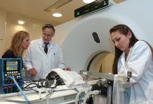
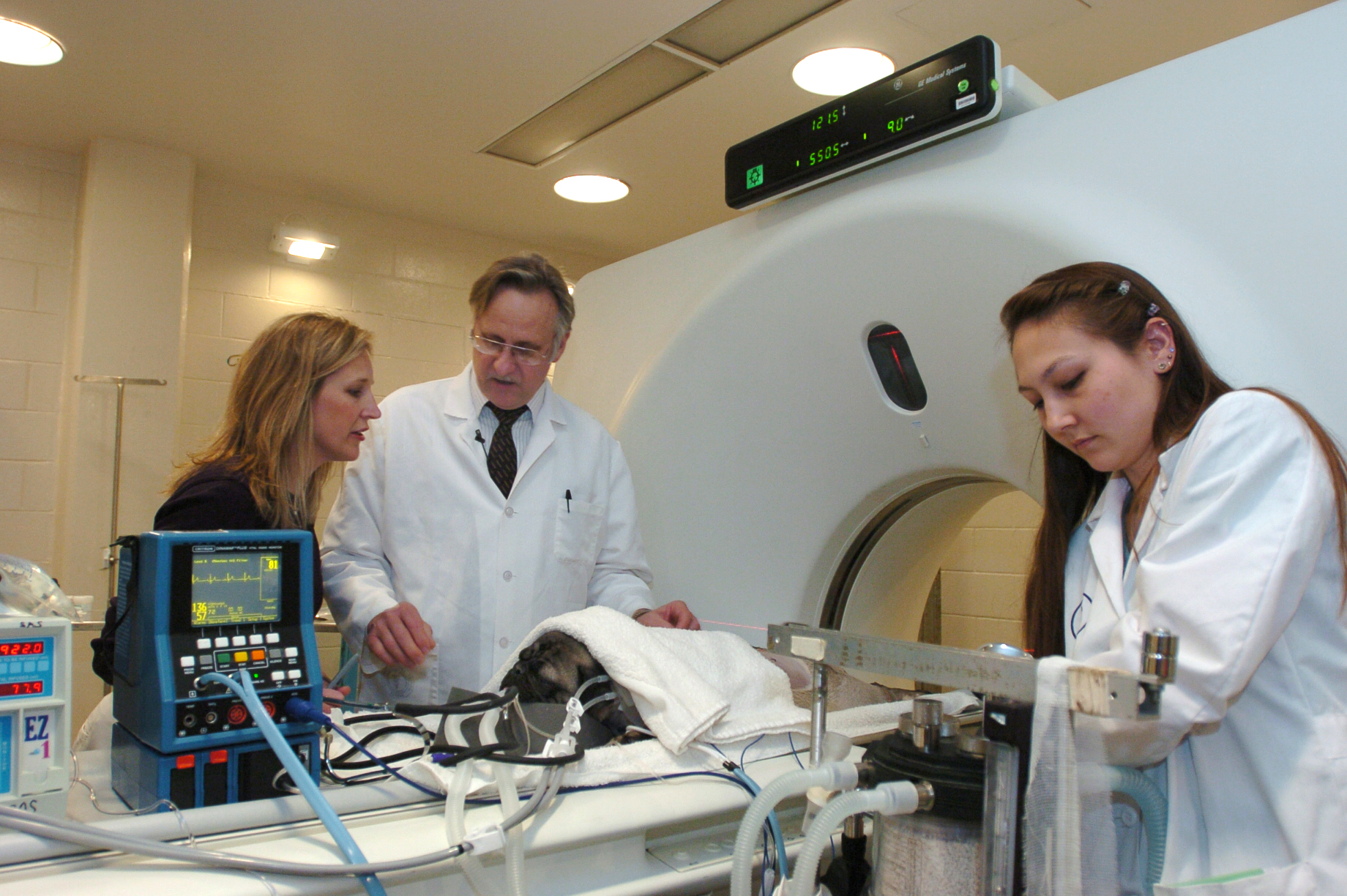
March 8, 2004 Purdue vet school gets state's first CT imager for animal patientsIndianapolis pug to receive CT assisted biopsyWEST LAFAYETTE, Ind. –Indiana's first CT scan for animals is now available at the Veterinary Teaching Hospital in Purdue University's School of Veterinary Medicine, offering digital imagery for diagnosis of internal injury and disease and decreasing the need for diagnostic surgery.
The refurbished $500,000 computed tomography imaging system will be used on Wednesday (3/10) to help diagnose the cause of pain in the lumbar vertebra of a 2-year old Indianapolis pug named Pug. The use of sophisticated diagnostic equipment is part of a national trend in veterinary medicine to provide health care similar to that available to humans. "The procedure should take 20 minutes or less for the scan and an additional 15 minutes for the biopsy," said William Blevins, professor and chief of diagnostic imaging in the Veterinary Teaching Hospital. "The case was referred by Dr. Derrell Elkins, a surgeon in Indianapolis. I initially read radiographs he took and gave my opinion on the diagnosis. The radiographs revealed that Pug has an osteomyelitis, or bone infection, of the fourth lumbar vertebra." Antibiotics have had no effect on the infection, so Blevins will perform the CT study to determine if there are any abscesses inside or adjacent to the vertebra that cannot be seen on regular radiographs. The CT imaging will also guide him during a biopsy of any tissue that appears to be abnormal. "A culture will be done following the procedure to determine the type of infection present and help us decide on an effective mode of treatment," Blevins said. The computer-based diagnostic machine, commonly used for human patients, creates detailed cross-sectional images of soft tissue and bone, which is helpful in locating and diagnosing internal injuries and conditions. The images can be reconstructed to make three-dimensional graphics that can aid in surgery and can be used to map the location of tumors for surgical removal and/or radiation therapy. "CT imaging has been in use in human medicine for many years, but is comparably new in veterinary medicine," Blevins said. "The ability to see inside our patients using the CT technology reduces the number of exploratory surgeries we have to perform and reduces the amount of time an animal has to spend in surgery under anesthesia. CT imaging also allows us to make a faster, more accurate diagnosis." Blevins said he is pleased with the results he and his staff have achieved in the last 10 months since the arrival of the CT imager. He is looking forward to the department's next first, a table being built in Wisconsin that will make it possible to obtain CT images of large animals, including horses weighing as much as 2,600 lbs. Blevins said the table will cost about $30,000, and he hopes it will be completed and installed in about two months. "With the special table, we will be able to image the limbs, head and neck of a horse, something we can’t do on the smaller table that was designed for human use," Blevins said. "Demand for the CT service is exceeding initial expectations, demonstrating the need for CT in diagnostics in many of the cases referred to the Veterinary Teaching Hospital by practitioners throughout Indiana and surrounding states," Blevins said. "The school was able to hold down costs by acquiring a refurbished CT machine that incorporates all the necessary modern technology. Additionally, by negotiating a special lease arrangement, we spread out the acquisition cost over a multiple year period." Writer: Reni Winter, (765) 496-3133, rwinter@purdue.edu Source: William Blevins, (765) 494-1122, blevinsw@purdue.edu Purdue News Service: (765) 494-2096; purduenews@purdue.edu Note to Journalists: Media wishing to attend the procedure can arrange to do so by contacting Reni Winter at (765) 496-3133. Related Web site: PHOTO CAPTION:
|
