Microsecond Raman imaging might probe cells, organs for disease
March 30, 2015
 |
|
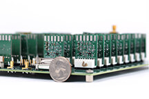
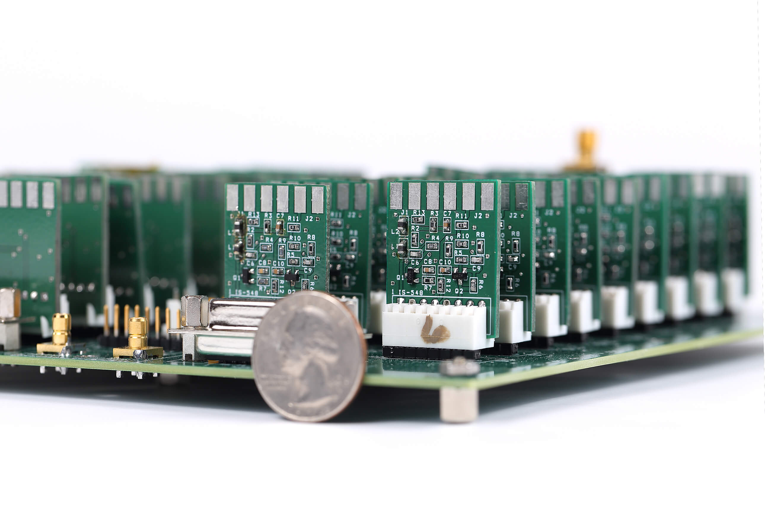
A new technology that could represent an advanced medical diagnostic tool for the early detection of cancer and other diseases is made possible with this electronic device developed at Purdue's Jonathan Amy Facility for Chemical Instrumentation in the Department of Chemistry. The device is called a 32-channel tuned amplifier array, or TAMP array. Two patents have been issued for the new technology. (Purdue University image) |
WEST LAFAYETTE, Ind. – A vibrational spectroscopic imaging technology that can take images of living cells could represent an advanced medical diagnostic tool for the early detection of cancer and other diseases.
High-speed spectroscopic imaging makes it possible to observe the quickly-changing metabolic processes inside living cells and to image large areas of tissue, making it possible to scan an entire organ.
"For example, we will be able to image the esophagus or urinary bladder for diagnosis of tumors," said Ji-Xin Cheng, a professor in Purdue University's Weldon School of Biomedical Engineering and Department of Chemistry. "If you were to take one millisecond per pixel, then it would take 10 minutes to obtain an image, and that's too slow to see what's happening in cells. Now we can take a complete scan in two seconds."
The technology represents a new way to use stimulated Raman scattering to perform microsecond-speed vibrational spectroscopic imaging, which can identify and track certain molecules by measuring their vibrational spectrum with a laser, a sort of spectral fingerprint.
Findings appeared on March 27 in the Nature Publishing Group journal Light: Science & Application.
The imaging technique is "label free," meaning it does not require samples to be marked with dyes, making it appealing for diagnostic applications. Another advantage of the new system is that it can be combined with another technique called flow cytometry to look at a million cells per second.
"You can look at large numbers of cells from a patient's blood sample to detect tumors, for example, and you can also look directly at organs with an endoscope," said Cheng, scientific director of the Label-free Imaging lab in the Birck Nanotechnology Center in Purdue's Discovery Park. "These capabilities will change how people use Raman spectroscopy for medicine. There are many organelles in each cell, and spectroscopy can tell us what's in the organelles, which is information not available by other techniques."
The paper was authored by Cheng; graduate students Chien-Sheng Liao, Junjie Li and Seung-Young Lee; research scientist Mikhail Slipchenko; Ping Wang, a post-doctoral research associate; and Robert Oglesbee, a former engineer at Purdue's Jonathan Amy Facility for Chemical Instrumentation in the Department of Chemistry.
As a proof of concept, the researchers demonstrated the new system by observing how human cancer cells metabolize vitamin A and how medications are distributed in the skin.
The technology, which is about 1,000 times faster than a state-of-the-art commercial Raman microscope, is made possible with an electronic device developed at the Jonathan Amy Facility called a 32-channel tuned amplifier array, or TAMP array. Two patents have been issued for the new technology. Cheng said the idea for this imaging technology was inspired by teaching undergraduates how the human ear amplifies sound. Circuits in the TAMP device do the same thing for optical signals, he said
Writer: Emil Venere, 765-494-4709, venere@purdue.edu
Sources: Ji-Xin Cheng, 765-494-4335, jcheng@purdue.edu
Note to Journalists: The research paper is available from the Nature Press Office at press@nature.com, or by contacting
Emil Venere, 765-494-4709, venere@purdue.edu
ABSTRACT
Microsecond Scale Vibrational Spectroscopic Imaging by Multiplex Stimulated Raman Scattering Microscopy
Chien-Sheng Liao#,1, Mikhail N. Slipchenko#,1, Ping Wang#,1, Junjie Li2, Seung-Young Lee1,
Robert A. Oglesbee3, Ji-Xin Cheng*1,3
1Weldon School of Biomedical Engineering, 2Department of Biological Sciences, 3Department of Chemistry, Purdue University
# equal contributions
*corresponding author, email: jcheng@purdue.edu, phone: +1 765-494-4335
Real-time vibrational spectroscopic imaging is desired for monitoring cellular states and cellular processes in a label-free manner. Raman spectroscopic imaging of highly dynamic systems is inhibited by relatively slow spectral acquisition on millisecond to second scale. Here, we report microsecond scale vibrational spectroscopic imaging by lock-in free parallel detection of spectrally dispersed stimulated Raman scattering signal. Using a homebuilt tuned amplifier array, our method enables Raman spectral acquisition, within the window defined by the broadband pulse, at the speed of 32 microseconds and with close to shot-noise limited detection sensitivity. Incorporated with multivariate curve resolution analysis, our platform allows compositional mapping of lipid droplets in single live cells, observation of intracellular retinoid metabolism, discrimination of fat droplets from protein-rich organelles in Caenorhabditis elegans, spectral detection of fast flowing tumor cells, and monitoring drug diffusion through skin tissue in vivo. The reported technique opens new opportunities for compositional analysis of cellular compartment in a microscope setting and high-throughput spectral profiling of single cells in a flow cytometer setting.
