
New imaging suites offer more diagnostic capabilities for animal health
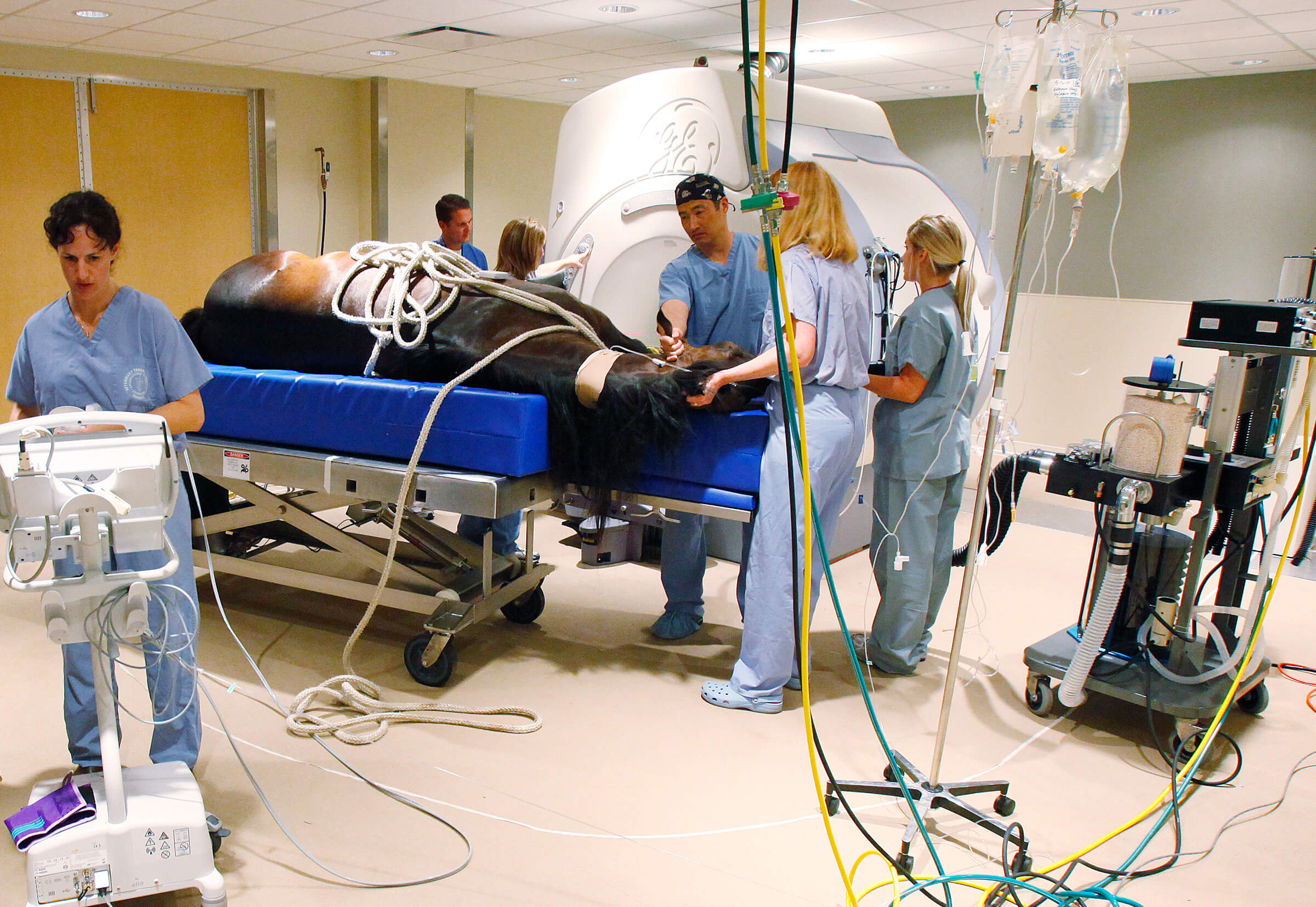
Heidi Lescun (far left), anesthesia technologist supervisor at the Purdue University Veterinary Teaching Hospital, checks an anesthesia monitor as other staff and faculty of the School of Veterinary Medicine prepare Wendy Jo, a quarter-horse, for an MRI in the new diagnostic imaging suites at the hospital. The horse was given the MRI to diagnose a suspected case of navicular disease, which causes pain in the foot.
WEST LAFAYETTE, Ind. - Veterinarians in the Purdue University School of Veterinary Medicine now have the ability to look for causes of lameness, neurological disorders and other conditions in horses thanks to new imaging equipment in the Veterinary Teaching Hospital.
"Purdue will be using the first equine-capable, high-field-strength, magnetic resonance imaging unit in Indiana," said Mimi Arighi, associate professor and director of the Veterinary Teaching Hospital. "The MRI is instrumental in diagnosing a variety of conditions, many of which cannot be detected using other forms of diagnostic imaging, in small and large animals. This machine and its new suite, as well as the new adjacent computed tomography suite, allows us to improve the care we provide animals."
The new suites are within the Veterinary Teaching Hospital building, which is located at 625 Harrison St. The MRI suite, which is more than 1,100 square feet, provides plenty of room to maneuver the patient and accommodate anesthesia equipment and personnel. Previously, patients had to be moved to a freestanding unit adjacent to the hospital.
"The MRI uses strong magnetic fields and radiofrequency pulse to produce images with superior soft tissue contrast and excellent detail," said James Naughton, a clinical assistant professor of radiology. "This imaging technology will facilitate the diagnosis of cancer and developmental and inflammatory disease of the brain, skull and spinal column in animals of all sizes. We also will be able to assess the extent of soft tissue disease and cancer for surgical and radiation therapy planning and treatment. It also will be used in the diagnosis of disease in the head and lower limbs of large animals, most commonly the horse."
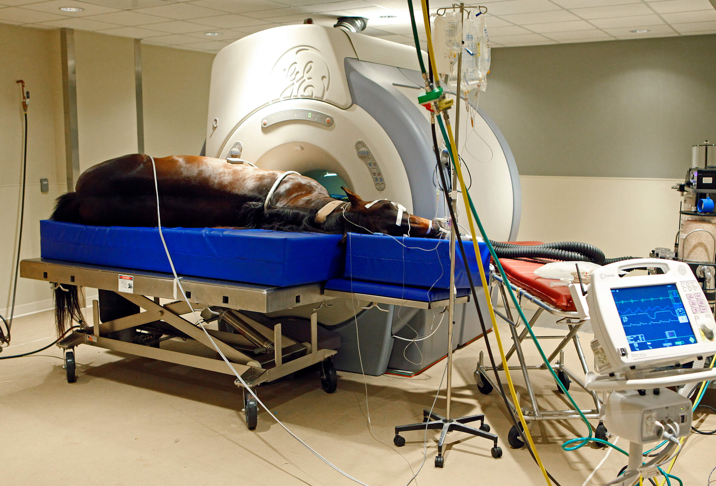
Quarter-horse Wendy Jo lies on an examination table during a break in an MRI in the new Purdue University Veterinary Teaching Hospital diagnostic imaging suites. The horse was being examined for a suspected case of navicular disease, which causes foot pain.
Currently the diagnostic imaging section performs about 17 small animal MRIs per month, and it is expected that four large animals will be scanned each month. Large animals receiving an MRI must be anesthetized and placed on a non-magnetic table that has been specially designed and built for animals that weigh more than 500 pounds. The same services can be offered for cattle, sheep, goats, pigs, llamas and alpacas. The GE 1.5 Tesla instrument is identical to those used in human applications and was paid for out of the hospital's operating budget.
The new multi-slice computed tomography unit, a recent donation from St. Elizabeth Regional Health, allows faster scanning of patients and increases the diagnostic information obtained during certain types of scans. It is one of only three such machines in Indiana, and more than 240 animals are scanned on the machine annually. This new suite is 707 square feet.
The CT unit, also commonly used for human patients, creates detailed cross-sectional images of soft tissue and bone, which is helpful in locating and diagnosing internal injuries and conditions. The images can be reconstructed to make three-dimensional graphics that can aid in surgery and can be used to map the location of tumors for surgical removal as well as radiation therapy.
Writer: Amy Patterson Neubert, 765-494-9723, apatterson@purdue.edu
James Naughton, 765-494-1107, jfnaught@purdue.edu
Related news release:
Purdue Veterinary School to install new CT scanner
Note to Journalists: Journalists interested in attending a media demo on Wednesday (May 12) at 1:30 p.m. should contact Kevin Doerr, director of alumni relations and public affairs for the