Projects on the Scanner
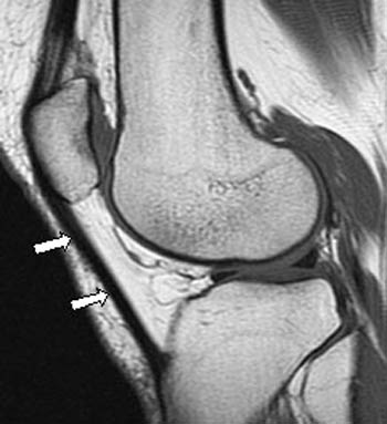 Arrows point out the patellar tendon, the main focus here.
Patellar Tendon Health: Diabetes
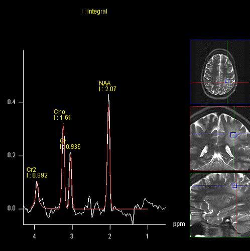 Spectra displayed of the human brain aid in detailed metabolic comparison.
Manganese Toxicity in Welders
PI: Ulrike Dydak
Operators: Humberto, Mahsa, Sandy Snyder
of Mn toxicity, which results in mood changes, cognitive deficits and eventually motor
symptoms similar to Parkinson’s disease. Our group is following a local cohort of welders since
2013 to study the dynamics of deposition of Mn in the brain via MRI, resulting changes in
neurochemistry via MRS, as well as correlations with cognitive and motor function, mood,
exposure metrics and more.
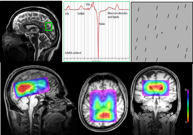 Top left:Single voxel GABA MRS voxel in the visual cortex; top middle: A typical GABA-edited spectrum; top right: visual stimuli for the subjects.
Bottom: A Novel 3D Multi-Voxel MRS Investigation of Autism using MEGA-LASER
Top left:Single voxel GABA MRS voxel in the visual cortex; top middle: A typical GABA-edited spectrum; top right: visual stimuli for the subjects.
Bottom: A Novel 3D Multi-Voxel MRS Investigation of Autism using MEGA-LASER
Attention Strengths and GABAergic Function in Children with Autism
Parsing the Stages of Reward Processing using Multimodal Neuroimaging
PI: Dan FotiOperators: Xiaopeng Zhou
Introduction: The brain's reward network is well-characterized, yet the time course of activity within this network during reward processing is poorly understood. This is critical insofar as reward processing dynamically unfolds across a series of functionally distinct stages, including: initial cue detection, motivated approach behavior, anticipation of outcome, and outcome evaluation. In this project, we will use a multimodal approach to characterize reward dynamics in a fine-grained manner, leveraging the spatial precision of fMRI and the temporal precision of event-related potentials (ERPs).
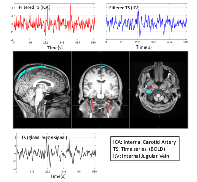
Understanding the Origin of Physiological Low Frequency Oscillations in BOLD fMRI Signal
PI: Yunjie TongIntroduction: Blood-oxygen-level dependent (BOLD) signal is the main contrast in fMRI to study brain function, however, there are physiological low frequency oscillations in BOLD with unknown origins. These low frequency oscillations are the confounding signals to the neuronal fluctuations. In this project, we will apply Real Time Phase-Contrast MRI to study the origins of these low frequency oscillations. We will target different blood vessels, from arteries to veins, to understand the dynamics of these nuisance signals and how they affect the neuronal fluctuation throughout the brain.
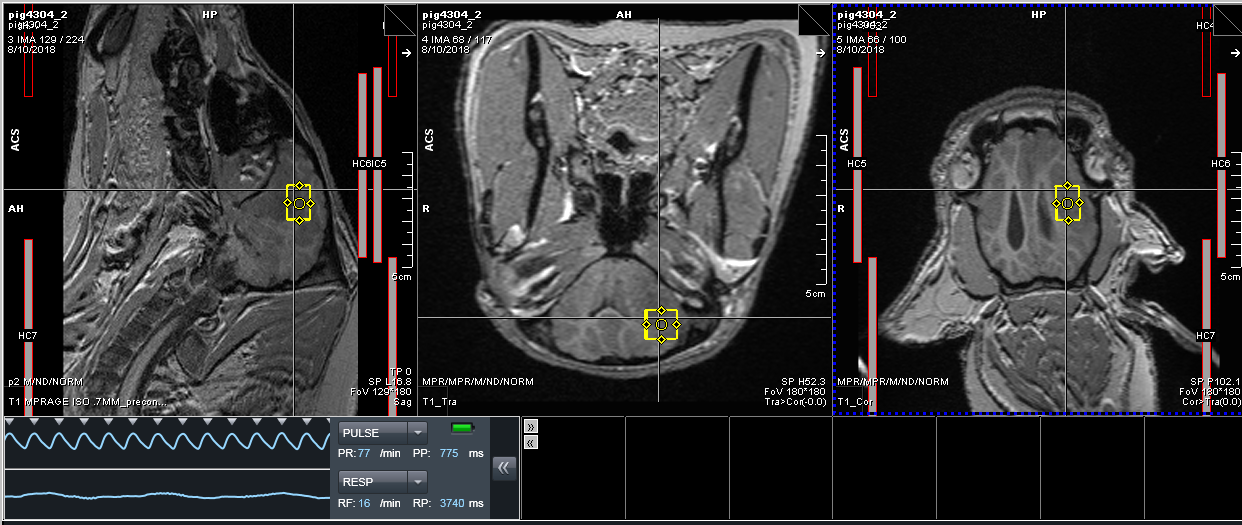
Development of a Swine Model to Evaluate Radiation-Induced Brain Injury: A Preliminary Report
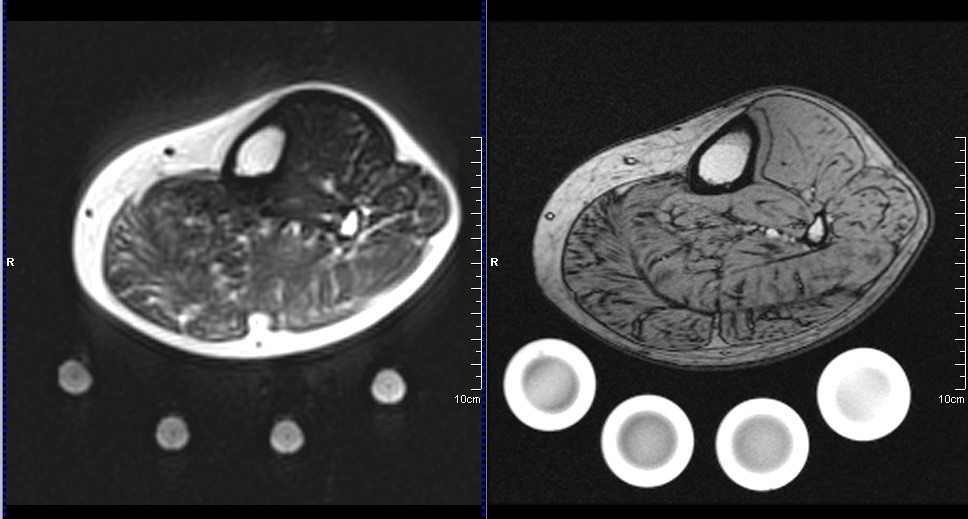
Musculoskeletal fat imaging and quantification by high-resolution metabolite cycling magnetic resonance spectroscopic imaging at 3 T: A fast method to generate separate distribution maps of lipid components
PI: Uzay Emir
Operators: Ahmad Alhulail, Pingyu Xia, Deb Patterson
Introduction: Current lipid evaluation techniques (Dixon and Single Voxel Spectroscopy) are unable to evaluate individual lipids or take too long. Biopsies provide this data, but are invasive. Therefore, there is a need for a reliable and fast non-invasive in vivo quantification method that is capable of providing the spatial distribution for each lipid of interest within a short scan time. In this work, we demonstrate a high-resolution, density-weighted concentric ring trajectory (DW-CRT) metabolite cycling (MC) MRSI acquisition technique to provide the needed fat quantification technique. The DW-CRT k-space-filling technique allows results of higher resolution within a shorter scan time. In addition, by implementing the MC acquisition technique, simultaneous out phased upfield and downfield (relative to the water frequency) spectra can be provided from a single acquisition. These spectra can be then used to reconstruct separate metabolites and water only spectra. By using this advantage, the water signal information can be used as an internal reference to calculate the FF voxel-wise in a similar approach used by the Dixon method, but for each lipid component individually based on their amplitude and unique resonance frequency. A second objective is to investigate the regional distribution of each lipid component over the calf muscles in an adolescent populatio