
Scanning Electron Microscope
- What is a SEM?
- How does a SEM work?
- How is a sample prepared?
- What are the radiation safety concerns?
- Scanning Electron Microscope Radiation Safety Guidelines
- References
What is a SEM?
SEM stands for scanning electron microscope. The SEM is a microscope that uses electrons instead of light to form an image. Since their development in the early 1950's, scanning electron microscopes have developed new areas of study in the medical and physical science communities. The SEM has allowed researchers to examine a much bigger variety of specimens.
The scanning electron microscope has many advantages over traditional microscopes. The SEM has a large depth of field, which allows more of a specimen to be in focus at one time. The SEM also has much higher resolution, so closely spaced specimens can be magnified at much higher levels. Because the SEM uses electromagnets rather than lenses, the researcher has much more control in the degree of magnification. All of these advantages, as well as the actual strikingly clear images, make the scanning electron microscope one of the most useful instruments in research today.
How does a SEM work?
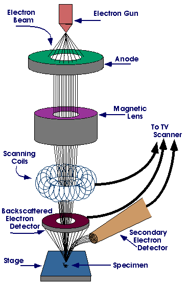
Diagram courtesy of Iowa State University
The SEM is an instrument that produces a largely magnified image by using electrons instead of light to form an image. A beam of electrons is produced at the top of the microscope by an electron gun. The electron beam follows a vertical path through the microscope, which is held within a vacuum. The beam travels through electromagnetic fields and lenses, which focus the beam down toward the sample. Once the beam hits the sample, electrons and X-rays are ejected from the sample.
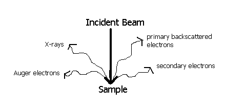
Detectors collect these X-rays, backscattered electrons, and secondary electrons and convert them into a signal that is sent to a screen similar to a television screen. This produces the final image.
How is a sample prepared?
Because the SEM utilizes vacuum conditions and uses electrons to form an image, special preparations must be done to the sample. All water must be removed from the samples because the water would vaporize in the vacuum. All metals are conductive and require no preparation before being used. All non-metals need to be made conductive by covering the sample with a thin layer of conductive material. This is done by using a device called a "sputter coater."
The sputter coater uses an electric field and argon gas. The sample is placed in a small chamber that is at a vacuum. Argon gas and an electric field cause an electron to be removed from the argon, making the atoms positively charged. The argon ions then become attracted to a negatively charged gold foil. The argon ions knock gold atoms from the surface of the gold foil. These gold atoms fall and settle onto the surface of the sample producing a thin gold coating.
What are the radiation safety concerns?
The radiation safety concerns are related to the electrons that are backscattered from the sample, as well as X-rays produced in the process. Most SEMs are extremely well shielded and do not produce exposure rates greater than background. However, scanning electron microscopes are radiation-generating devices and should be at least inventoried. The Indiana State Department of Health requires that the machines be registered with their office using State Form 16866, Radiation Machine Registration Application. It is also important that the integrity of the shielding is maintained, that all existing interlocks are functioning, and that workers are aware of radiation safety considerations.
The main reasons for developing a SEM safety plan are:
- to keep accurate inventory of all SEM's on campus (manufacturer/model, serial number, location, contact person and phone number)
- to warn workers of the risk of interfering with any safety devices (investigator needs to have permission to override any interlocks or warning devices)
- to make sure shielding is not compromised (exposure rate not greater than 0.5 mrem/hr at 5 cm from any surface of machine)
- to let workers know who to contact in an emergency or if they have any questions
Scanning Electron Microscope Radiation Safety Guidelines
- Safety evaluations will be performed initially when machine is purchased and after machine has been moved.
- Each machine should be key controlled when not in use. Interlocks, if present, must remain operational unless approved by the RSO.
- Shielding must be sufficient to maintain exposure rates less than 0.5 mrem/hr at 5 cm.
- The Radiation Safety Office will keep inventory and survey information on file in their offices. The SEM user should keep logbook of any maintenance done on machine. RSO must be notified if any modifications are made to the interlocks or any other safety devices. The SEM user should also keep a copy of operating and emergency procedures at the accelerator panel.
- No survey meters or personnel dosimetry are required.
References:
- Encyclopedia.Com
- Iowa State SEM Homepage
- Lawrence Livermore Radiation Safety Regulation, App. B, Summary of Radiation Generating Devices, Radiation Safety Requirements
- Virginia Tech Radiation Safety Pages
Many thanks also to the many responses and suggestions from the members of the RADSAFE list server.
If you have any questions please contact Radiological Management.
Campus Safety Contacts

If you see something, say something.
Purdue Police
Phone: (765) 494-8221
Purdue Fire
Phone: (765) 494-6919
Sign up for Emergency Text Messages
Do you want to recognize one of our staff for a job well done? Nominate the staff member(s) for a Bravo Award here!
