
Core Capabilities
The Purdue Imaging Facility (PIF) enables imaging of all types of biologic and non-biologic samples. Our systems can image samples ranging in size from rodents to nanometer sized objects. We train users how to use the imaging instruments and support users in sample preparation, anesthesia, image acquisition and analysis. The following paragraphs, Microscope Imaging and Pre-clinical and µCT, give an overview about our different imaging modalities.
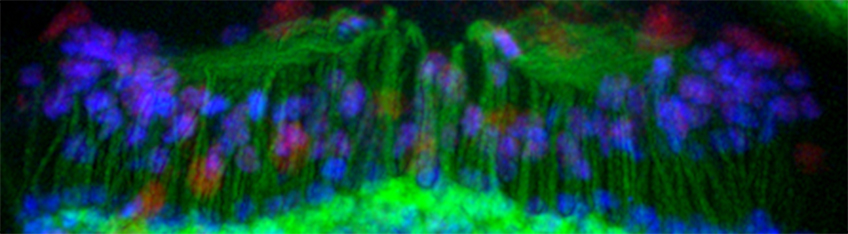
Microscope Imaging
Confocal Microscopy
Our facility provides three visible-wavelength laser point scanning confocal systems suitable for creating multi-dimensional images of living or non-living samples. Specimens labeled with single or multiple fluorophores as well as label-free samples (either transmitted or reflectance mode) can be visualized. Our confocal systems are built on both upright and inverted microscope platforms allowing for different sample preparations. All of our systems include motorized XY stages for large image stitching as well as repeating multi-xy positions during time-lapse imaging. The Nikon inverted confocals are equipped with a hybrid scanner system. It can be used for either high speed or high-resolution scanning as well as simultaneous imaging during Photo-activation experiments using the 405 nm laser to photo-activate and 488 nm/561 nm lasers to image photo-activation. In combination with a Tokai Hit live imaging stage top chamber and the perfect focus system, it is possible to collect short and long-term data of dynamic intercellular events at video frame rates while keeping the cells happy in 37oC and 5% CO2. The Zeiss confocal is built on an upright microscope. This allows the use of non-living/living samples under a coverslip or using unique objectives that are “dipped” directly into an aqueous solution the sample is placed thereby setting up an optically clean configuration. Regardless of the confocal for your application, all our systems are easy to use and provide excellent quality images. For additional detailed specifications, please contact the facility manager.
Multiphoton Microscopy
If your sample is thicker than 100 µm or you require less phototoxicity to your living sample, you might benefit by using a multi-photon (MP). We offer two MP systems that enable multi-dimension images of thick living or non-living samples. One system is based upon an inverted microscope while the other one is placed on an upright microscope both providing unique features. An important application where MP is utilized is live animal imaging, called Intravital Microscopy. We have the tools necessary to accommodate this application on our upright MP system. Multiphoton microscope can also be useful for photo-uncaging, photo-polymerization and other processes that require high-power, short wavelength light. For additional detailed specifications, please contact the facility manager.
Super Resolution & TIRF Microscopy
For spatial resolution beyond the diffraction-limit (<200 nm) we provide a microscope that perform two types of super resolution microscopy: Stochastic Optical Reconstruction Microscopy (STORM) and Structured Illumination Microscopy (SIM). Although both techniques use the same microscope, their operation is entirely different. STORM requires special fluorescent dyes and buffer and can achieve precision as high as 35 nm, typically on samples close to the coverslip. Our SIM system is compatible with common fluorescent dyes within the UV, green and mid-red excitation channels and enable resolution improvement around twice the diffraction limit. SIM is suitable for short live cell time lapse experiments. High-speed, multi-color TIRF is also possible for detecting signals very close to the coverslip allowing for dramatic increased signal to noise/background images. All these applications require a thin sample not to exceed ~7µm sitting under a #1.5 coverslip. For additional detailed specifications, please contact the facility manager.
Traditional Wide-field Modalities
We have 2 standard wide-field systems that include cameras as the detectors. We have an inverted microscope with a xenon light source equipped with standard blue, green, and red as well as far-red (700nm +) fluorescent filters and an EMCCD camera. It also has phase contrast, so we can acquire a transmitted image to show morphology of your cells/sample. Our second wide-field microscope is a simple upright microscope equipped with a very high-resolution camera for brightfield color imaging applications such as Immunohistochemistry. This system is also equipped with polarizers on both sides of the sample for capturing images using either Differential Interference Contrast or polarizing techniques like amyloid assays. For additional detailed specifications, please contact the facility manager.
Pre-clinical and µCT
The Bindley Imaging Facility provides systems capable of imaging larger subjects such as small rodents, medical devices or other non-biologics. Different modalities are available including: Optical Bioluminescence/Fluorescence and Computer Tomography (mCT) and imaging.
Spectral Ami Optical Imaging System
The SPECTRAL Ami optical imaging systems is an easy to use yet powerful in vivo imaging system but can also be used for non-living samples. This structural integrity ensures highly accurate and easily reproducible images and calibrations. It can perform either Fluorescent or Bioluminescent Imaging with a standard photography overlay. Software includes quantitative analysis features as well as multi-well template guide for measuring intensities on multi-well dishes. Our system is equipped with an anesthesia instrument to keep the animals still during scanning as well as a heated stage. Software can be downloaded on individual PCs to allow data viewing and quantification more conveniently than working at the PIF. For additional detailed specifications, please contact the facility manager.
Perkin Elmer Quantum GX µCT
The Quantum GX µCT apply low X-ray dose and enables true longitudinal imaging capability. It delivers high-resolution scanning for in vivo and ex vivo applications and is suitable for broad range of versatile small animal (from zebrafish to rabbits) application including: oncology, pulmonary and cardiovascular disease, diabetes, orthopedics and dentistry. Our system is equipped with an anesthesia instrument to keep the animals still during scanning as well as a software gating feature to help improve image quality. An advanced sub-volume reconstruction feature maintains a superior workflow and create high-resolution images from original whole image scan without the need to rescan samples. For additional detailed specifications, please contact the facility manager.
Other resources
Sample Preparation
Methods for preparing biological samples for high-resolution microscopic observation are almost as diverse as the samples themselves. Please follow the manufactures protocol for staining technique. A very good reference to start sample preparations would be the reference; Epi-Fluorescence Microscopy. Webb DJ, Brown CM. Methods in molecular biology (Clifton, N.J.). 2013; 931: 29-59. The following are some general considerations that are broadly applicable to many kinds of samples. Please always use a high quality #1.5 coverslip over your specimen. Also, consider using an anti-fade product to reduce photo-bleaching. They typically can be purchased from a vendor, such as Thermofisher, that act as both a mounting medium and anti-fade reagent. We do not always recommend these anti-fade reagents when supplied with the UV dye. For Super Resolution imaging or other questions pertaining to sample preparation, please contact the core director for more sample-specific advice.
Image Processing
Images may be analyzed qualitatively (e.g., by looking at them) or quantitatively. In either case, some post-processing is almost always desirable prior to analysis. Of course, post-processing alters the raw data, so the user must understand (and describe in a methods section!) all of the processing steps that were performed. We have several software platforms and offline workstations that can be used at no cost to conduct post-processing (such as blind deconvolution), optimization, and analysis on your image data to prepare for publication. Please contact the core manager for the appropriate workstation.
Contact
Xiaoguang (Shawn) Zhu
Imaging Facility Director
Phone: 765-496-3148
Email: zhu410@purdue.edu