

Purdue Imaging Facility
The Purdue Imaging Facility (PIF) is a research resource for Purdue faculty/staff/students and other academics and non-academics who want access to state-of-the-art light microscopy for their research and development work. PIF specializes in Confocal and Multi-Photon Microscopy, Pre-clinical Bioluminescence/Fluorescence and Computed Tomography. In addition, we are a resource for researchers at Purdue University and biotech companies around the community. PIF is a member of the Indiana CTSI service core community. We provide the instruments and expertise needed to visualize molecules in preparations ranging from single cells to entire animals. All facility users receive individualized instrument training as well as project-specific advice for optimal data acquisition. Consultation on sample preparation, image rendering, and data analysis are also available as our knowledge base permits. Collaborations and contract research services are encouraged.
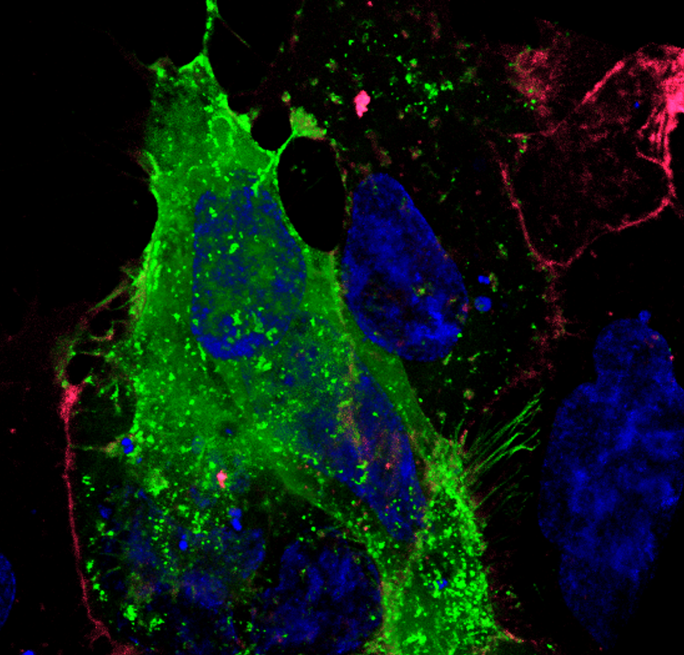
 Zeiss 880 confocal - breast cancer cells captured by Aparna Shinde
Common Applications
Confocal microscopy
Our facility provides three visible-wavelength laser point scanning confocal systems suitable for creating multi-dimensional images of living or non-living samples.
- Multi-colored fluorescence imaging
- Photoablation/photoactivation
- Imaging in aqueous environments
- Material science – fluorescence and reflectance
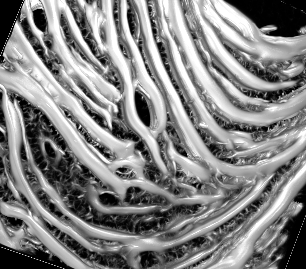 Nikon Intravital MP system - Second Harmonic Generation - collagen matrix captured by Jesus Hermida
Multiphoton microscopy
If your sample is thicker than 100 µm or you require less phototoxicity to your living sample, you might benefit by using a multi-photon (MP)
- Multi-dimensional images of thick samples, up to 800 microns
- Live cell and live animal (intravital) imaging
- Photo-uncaging
- Photoablation/photoactivation
- Second Harmonic Generation (non-centrosomatic)
 Nikon TIRF system - virus particles captured by Monica Husby
TIRF microscopy
Total Internal Reflection Fluorescence uses a finite wave-front of energy to fluorescently illuminate samples and limit excitation of fluorophores close to the coverslip to dramatically increase the signal to noise of your sample.
- Protein dynamic studies on the cell membrane
- Single molecule examinations
 Nikon SR system - Comparisons of Epi-Fluor vs Structured Illumination and STORM Micrograph captured by Andy Schaber
Super Resolution microscopy
For spatial resolution beyond the diffraction-limit (<220 nm) we offer Super Resolution microscopy with both SIM (3 colors) and STORM.
- Very high resolution intracellular imaging in thin samples
- Detailed localization analysis
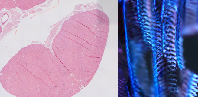 Nikon 90i system - Hematoxylin and Eosin and polarized illumination images captured by Andy Schaber
Wide-Field imaging
- Cell morphology
- Immunohistochemistry
- Polarization
- Amyloid assays
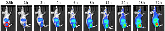 Spectral Instruments system - bioluminescence captured by Fanfei Meng
Pre-Clinical Molecular and microCT
The Bindley Imaging Facility provides systems capable of imaging larger subjects such as small rodents, medical devices or other non-biologics
- Small Animal CT Imaging
- Material science - Computed Tomography
- Fluorescent and Bioluminescent Imaging
 NIS Elements and Zen software platforms
Image Processing and analysis for all data
Images are great to display but have so much more data. Use one of our commerical analysis software tools to pull out all the information.
- Co-localization analysis
- Intensity analysis
- Time-lapse imaging of dynamic intracellular events
- Three-dimensional morphology
- Deconvolution
Contact
Xiaoguang (Shawn) Zhu
Imaging Facility Director
Phone: 765-496-3148
Email: zhu410@purdue.edu