 Purdue News
Purdue News
|
|
|
May 7, 2002 Lasers light way to 3-D imaging in Purdue labWEST LAFAYETTE, Ind. – Purdue University scientists developing a new imaging technology have created the world's first "visual fly-throughs" of a living tumor.
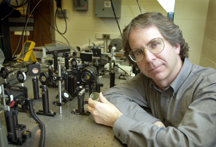
The technique, which uses lasers, holograms and special detectors, offers promise for a new kind of medical imaging based on light instead of tissue-damaging X-rays, said David Nolte, a professor of physics at Purdue. His research team recently used the new technique, called optical coherence imaging, to take a video of the insides of a cancerous rat tumor. "This is the first time that anybody has ever done a holographic fly-through of a living tumor," Nolte said. The tumor was not viewed while inside a rat, but was cultured and kept alive in a nutrient medium. Optical coherence imaging offers numerous possible applications, including diagnostic imaging for medicine and industry. The method might allow scientists to study how live tumors behave in real time, even how they react to experimental drugs. "This is a proof of concept," Nolte said. "These are the very first images of their kind." Findings about the work will be detailed in an oral paper to be presented May 23 during the Conference on Lasers and Electro-Optics in Long Beach, Calif. The paper will be presented by Ping Yu, a postdoctoral research associate at Purdue who authored the paper with Nolte, graduate student Mirela Mustata, John Turek, a professor of basic medical sciences, Michael Melloch, a professor of electrical and computer engineering, all from Purdue, and P.M.W. French, a physicist from the Imperial College of Science, Technology and Medicine in London. Critical to optical coherence imaging is a semiconductor holographic film developed by the team. The film is created out of a layered material produced by Michael Melloch, a professor of Electrical and Computer Engineering. Melloch uses a technique called molecular beam epitaxy to "grow" the material, layer by layer. Many other imaging technologies require that specimens, such as tumors, be specially prepared and cut into pieces for examination, killing the tissue. With optical coherence imaging, future medical researchers might use a joystick to interactively view living tissues, such as the internal structure of a tumor. "You can use a joystick to fly back and forth through a tumor," Nolte said. "If you see some structure worth looking at, you can back up, all in real time, and go section-by-section through the tumor." The new imaging technique is made possible by lasers and special "dynamic holographic films," – the most sensitive such films in the world – which have been developed in Nolte's lab since he came to Purdue a decade ago from Bell Labs. When two crossing laser beams are shined onto the film, holographic images are created. "This is holographic film that is adaptive, constantly changing and adjusting and moving in real time," Nolte said. "It's not a static hologram like you might see on the cover of a magazine. "These holograms adjust to the changing light conditions and the changing information that is carried on the laser beams. All of that coherent information is stored from the light so that it looks 3-D. It looks as if it were coming from the original object." The film is combined with a series of lenses and mirrors, acting as a filter that rejects ordinary, "scattered light" and lets through only the "coherent" laser light needed to produce the images. "I can take a flashlight in the dark, put it up against my hand, and my whole hand will glow red," Nolte said. "But I can't see any bones at all, even though the bones are just below the skin." The reason is because most of the light is said to be scattered, meaning it is a jumble of numerous separate light paths that do not move straight through the hand. "The emerging light is like ripples produced by a whole handful of rocks thrown at the same time into a pond," Nolte said. "The ripples run into each other, producing a chaotic mix of irregularly spaced waves." However, by using lasers, it is possible to find light paths that do go straight through an object. "In ordinary light, billions of atoms emit units of light called photons, and all of the photons are just in a random jumble," Nolte said. "A laser isn't like that. All of the atoms in a laser are communicating with each other, and they are all basically in lockstep." Lasers shine "coherent light" that could be likened to the ripples created by only a single rock thrown into a pond. The ripples, or light waves from a laser, are spaced regularly apart and move in unison. But most light detectors, including the human eye and conventional video cameras, do not detect coherent light, a limitation that can be overcome with the holographic film. The film is sensitive to coherent light. The holographic film in this case is made of alternating layers of two materials, gallium arsenide and aluminum gallium arsenide. These materials, semiconductors similar to those used to make lasers for CD players, form a 200-layer film. Each layer is 8 nanometers thick – or actually smaller than the wavelength of an electron in the material. Forcing electrons to move through layers so thin boosts the optical properties of the film and makes it more efficient at detecting coherent light. The Purdue team used the technique to take video images inside a tiny tumor, a form of cancer called osteogenic sarcoma, which afflicts bones and connective tissues. The tumor was about the size of a small pea. "One advantage of this method is that we can study tumors without ripping them apart," Nolte said. "That's important because when you rip them apart you kill them, which changes their physiology." The research is funded by the National Institutes of Health. The conference, known as CLEO, is considered one of the most important international gatherings for researchers in fields involving lasers and opto-electronics, which merges optics with electronics. CLEO is sponsored by the American Physical Society, the Optical Society of America, the Institute of Electrical and Electronics Engineers, and the Lasers & Electro-Optics Society. Nolte said he has been pleasantly surprised by the research findings. "There are all kinds of reasons why this should never have been possible and just a few reasons why, maybe, it was possible," he said. "But, of course, I never told my postdoc that. "And it worked." Writer: Emil Venere, (765) 494-4709, venere@purdue.edu Source: David Nolte, (765) 494-3013, nolte@physics.purdue.edu Purdue News Service: (765) 494-2096; purduenews@purdue.edu NOTE TO JOURNALISTS: A QuickTime animation showing the video "fly-throughs" also is available from Emil Venere, (765) 494-4709, venere@purdue.edu. PHOTO CAPTION: A publication-quality photograph is available at ftp://ftp.purdue.edu/pub/uns/nolte.imaging.jpeg. Video description:This animated graphic depicts a rat tumor. Purdue University researchers are developing a new imaging technology for creating the first "visual fly-throughs" of a living tumor. Unlike this image, which is static and shows sections of tissue that have been killed for imaging purposes, the fly-throughs enable scientists to see inside a living tumor in real time. (Purdue University Department of Physics)
ABSTRACT Visual fly-throughs of rat osteogenic sarcoma Dept. of Physics, Purdue University J.J. Turek Department of Basic Medical Sciences, School of Veterinary Medicine, Purdue University Michael Melloch School of Electrical and Computer Engineering, Purdue University P.M.W. French Femtosecond Optics Group, Physics Department, Imperial College of Science, Technology and Medicine We present the first depth-resolved images of living tumor tissue acquired directly to video camera using a coherence filter. The video fly-throughs reveal heterogeneous structure caused by necrosis and calcificatins characteristic of human tumors.
|
